Ultrasound Physics and Instrumentation
TLDRThis podcast explores ultrasound technology, detailing how to capture images, select transducers, and interpret artifacts. It traces the evolution from large-scale machines to portable devices, emphasizing the importance of ultrasound in point-of-care diagnostics and its role in reducing healthcare costs by minimizing unnecessary testing. The speaker delves into ultrasound physics, modes like B-mode, M-mode, and Doppler, and discusses the significance of frequency, body habitus, and transducer types. The presentation aims to equip listeners with the knowledge to perform effective ultrasound examinations.
Takeaways
- 🦇 Bats and dolphins have been using echolocation for thousands of years, and humans started using ultrasound in medicine in 1965.
- 📈 The evolution of ultrasound machines has transformed them from large, oscilloscope-connected devices to compact, handheld devices over the past 30 years.
- 🔍 Point-of-care ultrasound (POCUS) allows physicians to examine patients without the need for invasive procedures or ionizing radiation like CT scans.
- 🤝 Ultrasound enhances the physician-patient connection by enabling physicians to perform physical examinations and show patients their organ conditions in real-time.
- 📚 The 'Stanford 25' and 'UCI 30' are educational initiatives emphasizing the importance of physical examination skills, including the use of ultrasound, for medical students.
- 💡 Ultrasound works on the principle of sound wave reflection, timing the 'echo' to determine the depth and density of structures within the body.
- 🌟 Different ultrasound modes, such as B-mode, M-mode, and Doppler, serve various purposes, from imaging structure to assessing motion and blood flow.
- 🔧 Adjusting the ultrasound machine's settings, including depth, gain, and frequency, is crucial for optimizing image quality and penetration for different patient body types.
- 👁️ The choice of transducer affects the ultrasound's resolution and penetration; low-frequency transducers penetrate deeper but offer less resolution, while high-frequency transducers provide more detailed images but with less penetration.
- 🛠️ Understanding and managing artifacts like attenuation, enhancement, refraction, and reverberation is essential for accurate ultrasound interpretation.
- 🎯 Doppler ultrasound is vital for assessing blood flow, with pulse wave Doppler providing velocity waveforms and continuous wave Doppler detecting the highest velocities in stenotic conditions.
Q & A
What is the significance of ultrasound in medical imaging?
-Ultrasound is significant in medical imaging because it allows for real-time, non-invasive visualization of internal body structures without using ionizing radiation, which reduces the risk to patients.
How does ultrasound work according to the script?
-Ultrasound works by sending sound waves into the body. These waves travel through tissue and reflect back to the transducer. The time it takes for the echoes to return is used to create an image based on depth and density of the tissues.
What advancements have been made in ultrasound technology?
-Advancements in ultrasound technology include the reduction in size of the machines, making them more compact and portable, and the development of point-of-care ultrasound (POCUS) for bedside use.
What are some common modes used in ultrasound imaging?
-Common modes in ultrasound imaging include B-mode (brightness mode), M-mode (motion mode), color flow Doppler, and pulse wave Doppler. Each mode serves different purposes such as visualizing structures, assessing motion, and measuring blood flow.
What are some artifacts mentioned in the script and their significance?
-Artifacts such as posterior acoustic enhancement and reverberation artifacts are mentioned. These artifacts can help identify certain structures or indicate the presence of specific conditions, like gallstones or fluid collections.
How is depth adjusted in ultrasound imaging?
-Depth is adjusted using a control knob. Rotating it counterclockwise makes the depth shallower, while rotating it clockwise makes the depth deeper. This adjustment helps focus on the relevant area of interest on the screen.
What is the importance of transducer selection in ultrasound?
-Transducer selection is crucial as different transducers have varying frequencies and footprints, making them suitable for different types of examinations and depths of penetration. The right transducer provides the best resolution and image quality.
What role does Doppler play in ultrasound imaging?
-Doppler in ultrasound imaging is used to measure the flow of red blood cells in vessels, providing information about blood flow velocity and direction, which is essential for diagnosing vascular conditions and assessing cardiac function.
What is time gain compensation (TGC) and how is it used?
-Time gain compensation (TGC) is used to adjust the gain at different depths of the ultrasound image. It allows for uniform brightness across the image, compensating for the weakening of sound waves as they penetrate deeper into the body.
Why is patient data entry important in ultrasound examinations?
-Entering patient data is important to correctly archive images and ensure accurate diagnosis. It allows for proper tracking and association of ultrasound images with the respective patient, maintaining the integrity of medical records.
Outlines
🦇 Introduction to Ultrasound and its Evolution
The script introduces the topic of ultrasound, its instrumentation, and image acquisition. It discusses the use of ultrasound to obtain images, the types of transducers, and common artifacts. The history of ultrasound usage by animals like bats and dolphins, and later by humans in medicine since 1965, is highlighted. The evolution of ultrasound machines from large, oscilloscope-connected devices to compact, handheld ones is noted. The script emphasizes the importance of point-of-care ultrasound (POCUS) in connecting physicians with patients and its role in reducing healthcare costs by eliminating unnecessary testing and radiation exposure from procedures like CT scans.
🔬 How Ultrasound Works: Physics and Principles
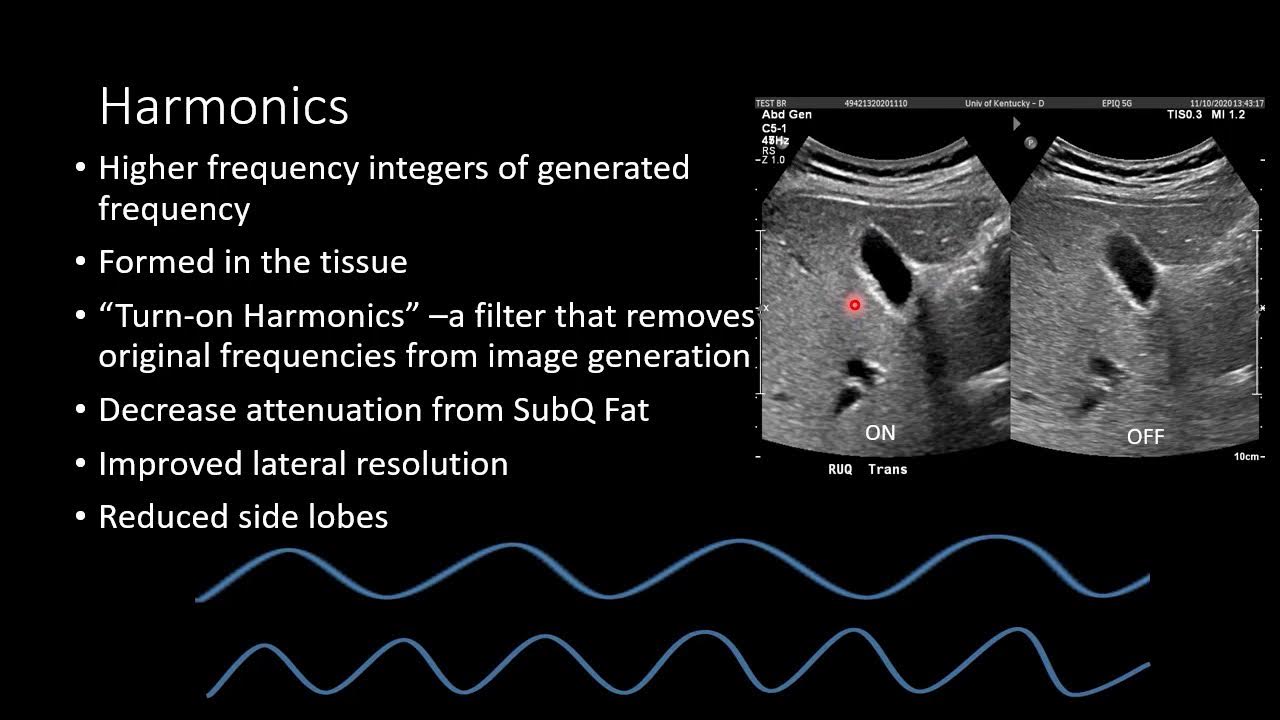
This paragraph delves into the physics behind ultrasound, comparing its operation to sonar with a reference to the movie 'The Hunt for Red October'. It explains how ultrasound uses the speed of sound traveling through human tissue to create images by emitting a pulse and measuring the time it takes for the echo to return. The concept of brightness mode (B-mode), motion mode (M-mode), and color flow Doppler (CF) are introduced, along with pulse wave Doppler (PW), to illustrate how they capture motion and flow within the body. The paragraph also discusses the importance of echogenicity, with examples of different densities in body structures and their appearance on ultrasound.
🔍 Ultrasound Transducers and Their Frequencies
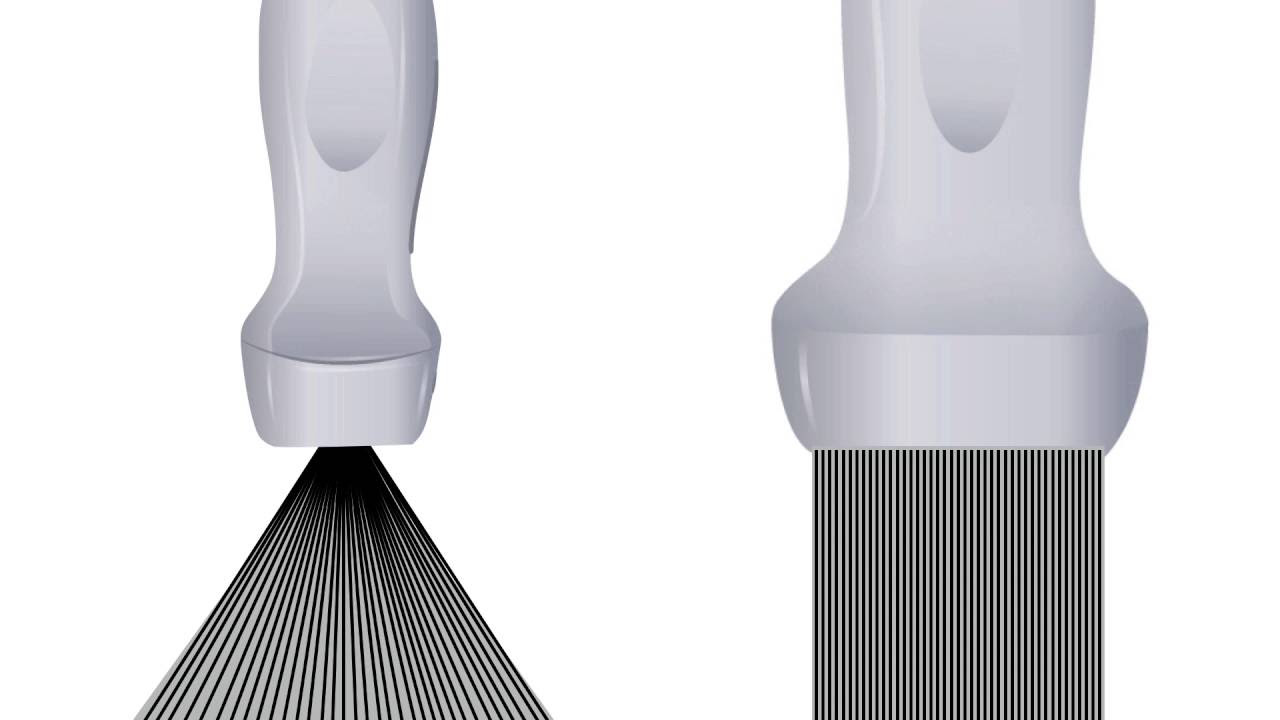
The script explains the importance of selecting the appropriate ultrasound transducer based on frequency and depth of penetration. It describes low, mid, and high-frequency transducers, comparing them to different camera lenses for their ability to see structures at varying distances. The paragraph details how to adjust frequency settings on a transducer and the impact of patient size on this choice. It also describes different types of transducers, such as curvilinear, phased array, and linear array, and their respective uses and imaging characteristics.
📏 Understanding Ultrasound Controls: Depth and Gain
This paragraph focuses on the significance of the depth control in ultrasound imaging, illustrating how adjusting depth can change the clarity and field of view of the image. It contrasts this with zoom, which magnifies a part of the screen without altering the image's fundamental resolution. The paragraph also discusses gain adjustments, including overall gain and time gain compensation (TGC), to balance the brightness and contrast of the ultrasound image at different depths.
🌐 Exploring Artifacts in Ultrasound Imaging
The script explores various artifacts that occur in ultrasound imaging, such as attenuation, posterior acoustic enhancement, and the challenges posed by gas in the body. It explains how these artifacts can be helpful in diagnosing conditions, such as differentiating a gallstone from a more serious issue. The paragraph also covers refraction, reverberation, mirror-image, and other artifacts, providing examples of how they appear on the ultrasound and their clinical significance.
🚀 Doppler Ultrasound: Flow Dynamics and Equations
This paragraph introduces Doppler ultrasound, explaining how it measures the flow of blood cells through vessels. It discusses the relationship between pressure, flow, and the radius of a vessel, emphasizing the impact of stenosis on flow velocity. The Doppler equation is introduced, highlighting the importance of the angle of insonation (theta) on Doppler accuracy. The paragraph also explains the difference between B-mode and Doppler in terms of beam angles and their effects on image quality.
🎛️ Adjusting Doppler Settings for Optimal Imaging
The script provides insights into adjusting Doppler settings on an ultrasound machine, such as pulse repetition frequency (PRF) and color gain, to optimize the Doppler signal. It discusses the trade-off between temporal resolution and Doppler sensitivity and the importance of being parallel to the flow for accurate Doppler measurements. The paragraph also explains the difference between pulse wave Doppler and continuous wave Doppler, and when to use each for optimal results.
📚 Mastering Ultrasound Controls and Patient Data Management
This paragraph concludes the script by emphasizing the importance of understanding ultrasound machine controls, such as depth, gain, frequency, and Doppler settings. It provides a brief overview of a specific ultrasound machine model, the GE Logic V2, and its control panel. The paragraph also stresses the importance of patient data management, including entering patient IDs and archiving images for accurate record-keeping and diagnosis.
Mindmap
Keywords
💡Ultrasound
💡Transducer
💡Artifacts
💡Point-of-Care Ultrasound (POCUS)
💡Ionizing Radiation
💡Doppler Effect
💡Echogenicity
💡Frequency
💡Transverse Plane
💡Sagittal Plane
💡Coronal View
Highlights
Ultrasound technology has evolved from large machines to compact handheld devices, facilitating point-of-care ultrasound (POCUS).
Bats and dolphins have used echolocation for thousands of years, and in 1965, humans started using ultrasound for medical imaging.
Ultrasound avoids the use of ionizing radiation, unlike CT scans, making it a safer diagnostic option.
The integration of ultrasound in physical examinations enhances the physician-patient connection and diagnostic accuracy.
The 'UCI 30' curriculum outlines 30 ultrasound skills medical students should master, covering the entire body.
Ultrasound can reduce healthcare costs by eliminating unnecessary testing and is a clinical re-engineering tool.
The physics of ultrasound is based on the time of flight of sound waves and their reflection to create images.
Different ultrasound modes, such as B-mode, M-mode, and Doppler, serve various diagnostic purposes.
Echogenicity is categorized into hyperechoic, hypoechoic, and anechoic to describe the reflection of sound waves.
Transducer frequency affects image resolution and penetration depth, requiring selection based on patient size.
Transducer footprints and types, such as phased array and linear array, are chosen based on the area of the body being examined.
The orientation of the ultrasound probe is crucial for accurate imaging and is guided by the probe's indicator.
Depth adjustment on the ultrasound machine is essential for optimizing image resolution and screen real estate.
Gain settings on the ultrasound machine affect the brightness of the image and need to be balanced for accurate diagnostics.
Artifacts such as attenuation and enhancement are not errors but valuable in identifying structures like gallstones.
Gas in the body can interfere with ultrasound imaging, and techniques like applying pressure can help overcome this.
Doppler ultrasound is essential for assessing blood flow, with different types like pulsed wave and continuous wave for various applications.
The use of Doppler is affected by the angle of insonation, with parallel alignment providing more accurate readings.
Temporal resolution in ultrasound is crucial for real-time imaging and is affected by the use of Doppler modes.
Annotating ultrasound images is vital for documentation and clarity, especially in complex or less obvious cases.
The GE Logic V2 ultrasound machine, introduced in 2016, offers a range of controls for optimizing diagnostic imaging.
Transcripts
Browse More Related Video
Basic Ultrasound Physics for EM
Ultrasound Modes, A, B and M Mode| Ultrasound Physics | Radiology Physics Course #12

Ultrasound Physics with Sononerds Unit 11

Doppler Ultrasound Part 1 - Principles (w/ focus on Spectral Waveforms)

Ultrasound Artifacts | Ultrasound Physics Course | Radiology Physics Course #25
Ultrasound Physics - Image Generation
5.0 / 5 (0 votes)
Thanks for rating: