Ultrasound Artifacts | Ultrasound Physics Course | Radiology Physics Course #25
TLDRThis educational video script delves into the world of ultrasound imaging, explaining the concept of artifacts as misrepresentations of anatomy. It explores the common B-mode ultrasound artifacts, such as edge shadowing, mirroring, reverberation, and others, caused by the ultrasound transducer's false assumptions about sound speed, travel path, and attenuation. The script offers insights into how these artifacts can be reduced and even used to deduce tissue types, emphasizing the importance of understanding the underlying physics for accurate ultrasound interpretation.
Takeaways
- 🌐 An ultrasound artifact is a misrepresentation of anatomy on the ultrasound image due to the machine's false assumptions about how sound behaves in tissues.
- 🔍 The ultrasound machine assumes sound travels at a constant speed (1541 m/s in soft tissue), but in reality, it varies with different tissues' properties.
- 📏 The machine also assumes sound travels in straight lines, but in reality, it can reflect and refract, leading to artifacts like edge shadowing and duplication.
- 🔄 Refraction artifacts occur when the ultrasound pulse hits a tissue boundary at an angle, causing a change in the angle of the ultrasound pulse and leading to edge shadowing or duplication of objects.
- 🔱 Mirroring artifacts happen when echoes reflect off the underside of an object and back to the transducer, creating a false mirror image on the opposite side of a highly reflective surface.
- 💥 Reverberation artifacts occur when echoes reflect back and forth between two highly reflective surfaces, creating multiple false echoes at equal spacings.
- 🌀 Ring down or Comet Tail artifacts are caused by fluid resonating at a set frequency, sending off echoes that create a ring down effect or a Comet Tail appearance.
- 📡 Side lobe or grating artifacts are due to the radial expansion of transducer elements, causing false echoes from highly reflective surfaces outside the main ultrasound beam.
- 🌑 Shadowing occurs when a highly attenuating or reflective structure prevents sound from passing through, resulting in no echoes behind the object and a dark area on the image.
- 🔊 Enhancement is the opposite of shadowing, where ultrasound pulses pass through a less attenuating object, resulting in a brighter area behind the object on the image.
- 🔄 Speed error artifacts are caused by the machine's assumption that sound speed is uniform in tissue, leading to incorrect plotting of tissue boundaries when sound speed varies.
- 🔄 Ambiguity artifacts occur when echoes from deeper structures return during the receive time for more superficial structures, causing ghosting or false plotting in the shallow field of view.
Q & A
What is an ultrasound artifact?
-An ultrasound artifact is a misrepresentation of the anatomy on an ultrasound image caused by the false assumptions made by the ultrasound transducer during the image creation process.
Why do artifacts occur in B mode ultrasound imaging?
-Artifacts occur in B mode ultrasound imaging due to the false assumptions made by the ultrasound machine, such as the constant speed of sound, straight-line travel of sound, and single reflection off tissue boundaries, which do not hold true in all cases.
What is the average speed of sound that ultrasound machines assume in soft tissue?
-Ultrasound machines assume that sound travels at an average speed of 1,514 meters per second in soft tissue.
What is edge shadowing and how does it occur?
-Edge shadowing is an artifact that occurs when the ultrasound pulse is refracted at an angle other than 90 degrees, causing a wedge of tissue to receive no ultrasound pulses and appear darker than it actually is.
How can the refraction artifact be reduced?
-The refraction artifact can be reduced by changing the angle of insonation, such as by angling the ultrasound transducer to achieve a more perpendicular interaction with the tissue surface.
What is mirroring in ultrasound imaging and what causes it?
-Mirroring is an artifact that occurs when an ultrasound pulse reflects off a highly reflective tissue boundary and then echoes off the underside of an object, creating a false duplicate of the object in the B mode image.
What is reverberation and how does it differ from mirroring?
-Reverberation is an artifact caused by an echo reflecting off the face of the ultrasound transducer, creating multiple false echoes at equal spacings. It differs from mirroring in that it involves the transducer itself rather than a tissue boundary.
What causes the ring down or Comet Tail artifact?
-The ring down or Comet Tail artifact occurs when fluid resonates at a set frequency due to the interaction with air around it, sending off resonant echoes towards the ultrasound transducer and creating a series of false reflections.
What is the side lobe or grating artifact and how does it occur?
-The side lobe or grating artifact occurs when sound energy from the side lobes of the ultrasound beam reflects off a highly reflective surface outside the main beam, sending back false echoes and causing incorrect plotting within the field of view.
What is shadowing in ultrasound imaging?
-Shadowing is a common artifact that occurs when an ultrasound beam encounters a highly attenuating or reflective structure, resulting in no ultrasound pulses passing through or beyond the structure, causing a dark area in the image.
What is enhancement and how can it help in identifying tissue types?
-Enhancement is an artifact that occurs when ultrasound pulses pass through a structure without significant attenuation, such as a fluid-filled structure, and appear with higher intensity than expected. It can help in identifying tissue types by revealing the presence of fluid-filled structures as opposed to highly reflective ones like air.
What is ambiguity in ultrasound imaging and how does it occur?
-Ambiguity is an artifact that occurs when returning echoes from deeper structures are plotted within a shallower field of view due to a reduced pulse repetition period, causing a ghosting effect of the object in the shallower depth.
Outlines
🔬 Understanding Ultrasound Artifacts
Introduction to the concept of ultrasound artifacts. Artifacts are misrepresentations of the anatomy on ultrasound images. The paragraph revisits Doppler ultrasound imaging and aliasing, explaining how incorrect calculations lead to artifacts. It sets the stage for discussing B mode ultrasound artifacts, which arise from the assumptions made by the ultrasound transducer.
🚀 Key Assumptions Leading to Artifacts
Explains the false assumptions made by ultrasound machines that result in artifacts. These include sound traveling at a constant speed, in straight lines, and reflecting off tissues once. It also covers the idea that echoes return from a perpendicular surface and the assumption of linear attenuation of sound, highlighting how deviations from these assumptions create artifacts.
🔄 Refraction Artifacts Explained
Discusses refraction artifacts in ultrasound imaging. Refraction occurs when ultrasound pulses enter tissues at non-perpendicular angles, causing changes in the transmission angle. This results in edge shadowing and incorrect plotting of objects due to angle changes. Solutions to mitigate these artifacts are also discussed, like changing the angle of insonation.
🌟 Mirroring and Reverberation Artifacts
Details mirroring artifacts, which occur due to highly reflective tissue boundaries creating mirror images of objects. It also explains reverberation artifacts, where multiple echoes between strong reflectors create repeated, false images. Both types illustrate how reflections and echo paths lead to incorrect representations in ultrasound images.
✨ Side Lobe and Shadowing Artifacts
Explains side lobe artifacts caused by reflections from outside the primary ultrasound beam, leading to false echoes within the field of view. Shadowing artifacts occur when ultrasound beams encounter highly attenuating or reflective structures, causing regions with no returning echoes. The discussion includes examples like ribs and air bubbles.
💡 Acoustic Enhancement and Speed Errors
Covers acoustic enhancement artifacts where areas behind low-attenuating structures appear brighter due to less sound attenuation. Speed error artifacts occur when sound travels at different speeds through various tissues, causing incorrect plotting of tissue boundaries. Examples include the effect of fat and muscle on ultrasound speed.
⏳ Understanding Ambiguity Artifacts
Describes ambiguity artifacts related to the pulse repetition period (PRP) in ultrasound imaging. Short PRPs, used for superficial imaging, can result in echoes from deeper structures being incorrectly plotted within the shallower field of view, creating ghost images. This section explains how adjusting PRP impacts imaging depth and artifact occurrence.
📚 Preparing for Ultrasound Physics Exams
Concludes with a reminder of the importance of understanding artifacts for ultrasound physics exams. It introduces a curated question bank with video explanations for common exam questions, encouraging viewers to use this resource for study. The final video of the course on ultrasound safety is also mentioned.
Mindmap
Keywords
💡Artifact
💡Assumption
💡Refraction
💡Edge Shadowing
💡Mirroring
💡Reverberation
💡Ring Down or Comet Tail Artifact
💡Side Lobe or Grating Artifact
💡Shadowing
💡Acoustic Enhancement
💡Ambiguity
Highlights
An ultrasound artifact is a misrepresentation of anatomy on the ultrasound image.
Artifacts occur due to false assumptions made by the ultrasound transducer during image creation.
The assumption of constant sound speed in tissues leads to artifacts, despite varying speeds in reality.
The misconception that sound travels in straight lines can cause artifacts due to reflections and refractions.
Ultrasound machines assume echoes return from perpendicular surfaces, which is not always the case.
The assumption of single reflections off tissue boundaries can result in multiple echo artifacts.
Linear attenuation of sound is assumed by the transducer, but attenuation varies with tissue types.
Edge shadowing artifact occurs due to refraction of sound waves at the edges of objects.
Refraction can also cause duplication artifacts when echoes are incorrectly plotted.
Mirroring artifact is caused by echoes bouncing off the underside of highly reflective surfaces.
Reverberation artifact happens when echoes reflect off the ultrasound transducer face.
Ring down or Comet Tail artifact is caused by resonant echoes in fluid between microbubbles.
Side lobe or grating artifact results from radial expansion of transducer elements.
Shadowing occurs when ultrasound waves cannot penetrate highly attenuating or reflective structures.
Acoustic enhancement appears behind objects that do not strongly attenuate ultrasound.
Speed error artifacts are caused by the incorrect assumption of uniform sound speed in tissues.
Ambiguity artifacts happen when returning pulses from deeper structures are plotted in a shallower field of view.
Understanding artifacts is crucial for ultrasound exams as it tests knowledge of underlying physics.
A curated question bank of ultrasound questions from various Radiology physics exams is provided for study.
Transcripts
Browse More Related Video

Unit 21: Acoustic Artifacts

Ultrasound Physics Review | Velocity Error Artifacts | Sonography Minutes

Ultrasound Physics Review | What Are Artifacts | Sonography Minutes
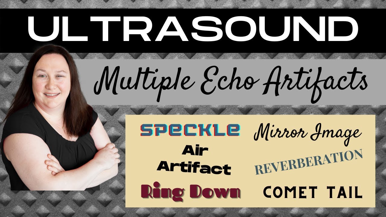
Ultrasound Physics Review | Multiple Echo Artifacts | Sonography Minutes

Ultrasound Physics Review | Beam Artifacts | Sonography Minutes
Time Gain Compensation | Ultrasound Physics | Radiology Physics Course #13
5.0 / 5 (0 votes)
Thanks for rating: