Ultrasound Physics - Image Generation
TLDRThis screencast delves into the physics of ultrasound imaging, explaining how mechanical energy in the form of ultrasound waves creates images by reflecting off tissue interfaces. It covers the impact of transducers on image quality, the role of acoustic impedance, and the use of high and low frequency probes for different imaging needs. The video also introduces advanced concepts like harmonics and compound imaging, which enhance image resolution and clarity, while minimizing artifacts.
Takeaways
- 🌌 Ultrasound waves are a form of mechanical energy that cause compression and rarefaction in tissues as they pass through.
- 🔍 The frequency range for medical imaging ultrasound waves is about 1 to 20 megahertz, with an average soft tissue velocity of 1540 meters per second.
- 🛡 Reflection of ultrasound waves off tissue interfaces is crucial for image production, while scattering, refraction, and absorption of waves can reduce signal quality and create noise and artifacts.
- 📐 Acoustic impedance, the product of the speed of sound and tissue density, affects the strength of wave reflection and is a key factor in image quality.
- 🔄 Harmonic imaging improves image quality by filtering out the original frequency and listening only for the higher frequency harmonics created by tissue resonance.
- 🔄 Compound imaging enhances image quality by generating multiple pulses at different angles and averaging them to reduce noise and accentuate high-level reflectors.
- 🛠 Piezoelectric crystals in the ultrasound probe generate and detect ultrasound waves, converting electrical energy to mechanical energy and vice versa.
- 🔄 Axial resolution in ultrasound imaging is improved by increasing frequency and decreasing wavelength, allowing better discrimination between tissue interfaces.
- 📏 Lateral resolution is influenced by the density of ultrasound waves in the field of view, with higher density leading to better resolution.
- 📈 Elevation resolution, akin to slice thickness, affects how much tissue is averaged to generate a 2D image, with thinner slices providing less averaging and potentially better resolution.
- 🔧 Different probe types, such as phased array, linear array, and curved probes, are used for different imaging needs based on factors like resolution, penetration, and field of view.
Q & A
What is the primary form of energy used in ultrasound imaging?
-The primary form of energy used in ultrasound imaging is mechanical energy, which is generated by the piezoelectric crystals within the probe and transmitted as ultrasound waves into the tissue.
What is the frequency range of ultrasound waves used in medical imaging?
-The frequency range of ultrasound waves used in medical imaging is approximately 1 to 20 megahertz.
How does the velocity of ultrasound waves change as they pass through different tissue types?
-The velocity of ultrasound waves changes based on the tissue types they pass through, with the velocity being highest in bone, slower in soft tissue, even slower in fluid, and slowest in gas.
What is the significance of the reflection of ultrasound waves in image production?
-The reflection of ultrasound waves off of tissue interfaces is significant in image production, as these reflected waves are what produce the image. The strength of the reflection is based on factors such as acoustic impedance, surface smoothness, tissue interface size, and orientation to the probe.
What is meant by the term 'acoustic impedance' in the context of ultrasound imaging?
-Acoustic impedance is a physical principle defined as the product of the speed of sound in a tissue and the tissue's density. It is important because interfaces between two tissues with different acoustic impedances generate echoes, which contribute to the ultrasound image.
How does the orientation of a tissue interface to the ultrasound probe affect image quality?
-The orientation of a tissue interface to the ultrasound probe affects image quality because if the interface is oriented perpendicular to the probe, it will act as a strong reflector and produce a clear image. If it is oriented parallel, it will be a poor reflector and result in reduced image quality.
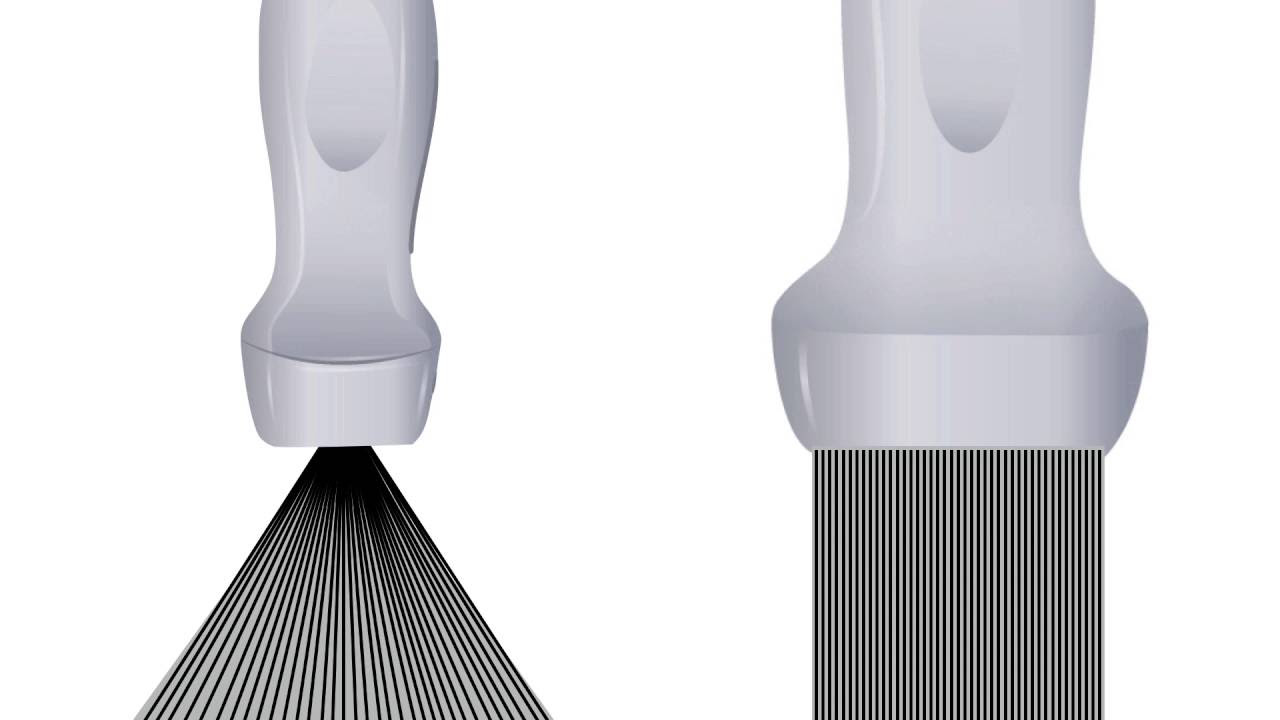
What is the difference between a phased array probe and a linear array probe in terms of imaging capabilities?
-A phased array probe has multiple crystals along the surface of the transducer and is typically lower frequency, allowing for better penetration but lower resolution. A linear array probe has crystals that are sequentially excited, resulting in higher resolution and is often used for imaging superficial structures.
What is the concept of 'harmonics' in ultrasound imaging and how does it improve image quality?
-Harmonics in ultrasound imaging refers to the new ultrasound waves generated at a frequency that is an integer multiple of the original frequency due to tissue resonance. By listening only for these harmonic frequencies, the ultrasound machine can reduce attenuation and improve lateral resolution, leading to a crisper and higher resolution image.
What is compound imaging and how does it affect the ultrasound image?
-Compound imaging is a technique where multiple pulses are generated at different angles within the same field of view, and the images from each pulse are averaged to create the final image. This reduces noise and accentuates high-level reflectors, resulting in a clearer image but at the cost of a lower sweep speed.
How does the concept of 'elevation resolution' relate to the thickness of the ultrasound image?
-Elevation resolution is related to the thickness of the ultrasound image in that a thicker slice results in more tissue being averaged together to generate a 2D image, while a thinner slice results in less tissue being averaged, leading to a more detailed and precise image.
What are the trade-offs between using high frequency probes and low frequency probes in ultrasound imaging?
-High frequency probes provide high resolution images but have lower penetration, making them suitable for imaging superficial structures. Low frequency probes offer high penetration at the expense of resolution, which is useful when imaging deeper tissues.
Outlines
🌀 Understanding Ultrasound Image Creation
This paragraph delves into the physics of ultrasound imaging, explaining how mechanical energy in the form of ultrasound waves is used to create images. The waves cause compression and rarefaction of tissues, with reflections from tissue interfaces generating the image. Factors like acoustic impedance, tissue interface characteristics, and orientation affect the strength of these reflections. The paragraph also touches on wave behavior, including scattering, refraction, and absorption, which can introduce noise and artifacts into the image.
🔊 Piezoelectric Crystals and Image Resolution
The second paragraph focuses on the generation of ultrasound pulses using piezoelectric crystals and how they impact image resolution. It explains the concepts of axial, lateral, and elevation resolution, detailing how the characteristics of the ultrasound pulse, including frequency and wavelength, determine the clarity of the image. The paragraph also introduces different types of probes, such as phased array and linear array, each with their advantages and trade-offs between resolution and penetration depth.
🔍 Probe Types and Advanced Imaging Techniques
This section discusses various probe types, including phased array, linear array, and curved probes, highlighting their specific uses and how they contribute to imaging different body parts. It also introduces advanced imaging techniques like harmonics and compound imaging. Harmonics improve image quality by filtering out lower frequency signals, leading to better resolution and reduced artifacts. Compound imaging, on the other hand, involves taking multiple pulses at different angles and averaging them to reduce noise and enhance image clarity, although it may also reduce real-time imaging speed.
🛠️ Reflectivity, Image Quality, and Diagnostic Artifacts
The final paragraph summarizes the key factors that influence the reflectivity of ultrasound waves, such as tissue interfaces and acoustic impedance differences. It emphasizes the importance of surface smoothness and perpendicular orientation to the probe for optimal reflection. The paragraph also discusses the use of high and low-frequency probes for different imaging depths and resolutions. Additionally, it explains how harmonics and compound imaging can be adjusted to accentuate or reduce diagnostic artifacts, providing a clearer understanding of the image quality and its impact on medical diagnosis.
Mindmap
Keywords
💡Ultrasound Image
💡Transducers
💡Acoustic Impedance
💡Reflection
💡Scattering
💡Refraction
💡Absorption
💡Piezoelectric Crystals
💡Resolution
💡Phased Array
💡Harmonics
💡Compound Imaging
Highlights
Ultrasound image generation involves the use of mechanical energy in the form of ultrasound waves that cause tissue compression and rarefaction.
Ultrasound waves used in medical imaging have a frequency range of 1 to 20 megahertz and travel at different velocities depending on the tissue type.
Reflection of ultrasound waves off tissue interfaces is the primary mechanism for image production, while other interactions like scattering, refraction, and absorption can reduce signal quality.
Acoustic impedance, the product of the speed of sound and tissue density, plays a crucial role in the strength of wave reflection and image clarity.
Optimal reflectors in ultrasound imaging are smooth, perpendicular to the sound waves, and have a high acoustic impedance difference between adjacent tissues.
Scattering occurs on irregular surfaces and results in a loss of signal, impacting image generation negatively.
Refraction can cause wave angulation and signal intensity reduction, leading to image artifacts.
Absorption of ultrasound waves converts mechanical energy into thermal energy, affecting the signal within the image and potentially causing artifacts.
Ultrasound pulses are generated using piezoelectric crystals that resonate at specific frequencies to create mechanical energy waves.
Image resolution in ultrasound is influenced by the characteristics of the pulse, including the number of sound waves generated and their wavelength and frequency.
Axial resolution improves with shorter wavelengths and higher frequencies, allowing better discrimination between tissue interfaces.
Lateral resolution is influenced by the density of ultrasound waves in the field of view, with higher density leading to better discrimination.
Elevation resolution relates to the slice thickness in ultrasound imaging, affecting the amount of tissue averaged to generate the 2D image.
Phased array probes use multiple crystals to generate a sector image with lower frequency for better penetration but lower resolution.
Linear array probes provide high-resolution imaging of superficial structures through sequential excitation of crystals.
Curved probes, or curved linear probes, offer a wide field of view with varying degrees of curvature for different imaging applications.
Harmonic imaging improves image quality by filtering out the original frequency and listening only for the harmonic frequencies generated through tissue resonance.
Compound imaging reduces noise and enhances image quality by generating multiple pulses at different angles and averaging the reflections.
The choice between high and low frequency probes depends on the need for resolution versus penetration in ultrasound imaging.
Harmonics and compound imaging are standard features on most ultrasound machines, improving image quality but can sometimes be adjusted to accentuate diagnostic artifacts.
Transcripts
Browse More Related Video

Clarius: Fundamentals of Ultrasound 1 (Physics)
Basic Ultrasound Physics for EM
Time Gain Compensation | Ultrasound Physics | Radiology Physics Course #13

Ultrasound Probes and Transducer Types | Ultrasound Physics | Radiology Physics Course #14

Ultrasound Physics with Sononerds Unit 12a

Unit 21: Acoustic Artifacts
5.0 / 5 (0 votes)
Thanks for rating: