Pregnancy - How a Wonder is Born! (Animation)
TLDRThe transcript details the remarkable journey of human reproduction and development, starting from the ovary's egg follicles to the complex process of fertilization. It describes the intricate dance of sperm and egg, leading to the formation of a zygote and its evolution into a fetus. The narrative unfolds the stages of embryonic growth, highlighting key milestones such as neuralation and the development of organs, culminating in the fully-formed fetus ready for birth after approximately 38 weeks.
Takeaways
- 🌟 The human reproductive system is equipped with specialized organs for creating new life, including the ovaries with about half a million eggs.
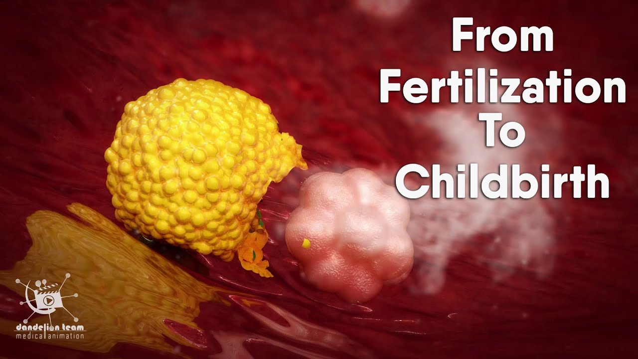
- 🥚 Ovulation involves the release of an egg, which is protected by the corona radiata and zona pellucida layers, essential for sperm penetration.
- 🧬 The egg cell contains 23 chromosomes with DNA that holds the blueprint for body cells, traits, and functions.
- 💫 Sperm cells have a single mission to reach and fertilize the egg, guided by messenger substances emitted by the egg itself.
- 🌱 Fertilization results in the formation of a zygote, the first cell of a new human being, which undergoes a series of cell divisions.
- 🍇 The early embryo, called a morula, and later a blastocyst, is nourished and protected by the zona pellucida until implantation in the uterus.
- 🚨 Implantation is a critical process where the blastocyst hatches from the zona pellucida and embeds itself into the uterine lining.
- 🔄 Gastrulation leads to the formation of three germ layers (ectoderm, mesoderm, and endoderm) that will develop into various tissues and organs.
- 🧠 By the third week of development, neuralation occurs, laying the foundation for the central nervous system, including the brain and spinal cord.
- 👶 By the ninth week, the embryo becomes a fetus with a human-like appearance, and further development includes the growth of features like eyelids, limbs, and facial structures.
- 📈 The fetus grows and matures, receiving oxygen and nutrients through the placenta and umbilical cord, while waste products are transferred to the mother's blood for excretion.
Q & A
How many eggs does the ovary possess?
-The ovary possesses about half a million eggs embedded in follicles.
What are the two protective layers surrounding the egg cell?
-The two protective layers surrounding the egg cell are the corona radiata and the zona pellucida.
What is the role of DNA in the formation of body cells?
-DNA contains the blueprint for building our body cells, determining eye color, body size, arrangement, and function of our organs.
How is the egg cell transported inside the fallopian tube?
-The egg cell is transported inside the fallopian tube with the help of cilia and mucus produced by glands in the walls of the fallopian tube, along with additional contractions of the muscle layers.
What happens when a sperm cell penetrates the protective layers of the egg?
-Upon penetration, the acrosome cap of the sperm cell degrades, leading to fertilization. This activation causes changes in the zona pellucida to prevent further sperm penetration and triggers the egg to complete its second maturation division.
What is the result of the fertilization process?
-Fertilization results in the formation of a zygote, which is the very first cell of the new human being, containing a combination of the mother's and father's genetic information.
How does the blastocyst prepare for implantation in the uterus?
-The blastocyst hatches from the protective zona pellucida and then burrows further into the uterine lining, much like a parasite, to prepare for implantation.
What are the three germ layers formed during gastrulation?
-The three germ layers formed during gastrulation are the ectoderm, mesoderm, and endoderm, which eventually differentiate into various tissues and organs.
What is the function of the placenta and umbilical cord during fetal development?
-The placenta and umbilical cord facilitate the exchange of substances between the mother and fetus, providing oxygen, nutrients, and removing waste products.
At what stage does the fetus achieve a human-like appearance?
-The fetus achieves a human-like appearance during the ninth week and in the third month of development.
What are the key milestones of the fourth week of development?
-In the fourth week of development, the central nervous system is established with the formation of the neural tube, the heart begins pumping blood, and limb buds start to appear.
Outlines
🌟 Fertilization and Early Development
This paragraph describes the process of fertilization and the initial stages of human development. It begins with the mention of the reproductive organs of men and women, specifically the ovary and its half a million eggs. The dominant follicle's maturation and ovulation are detailed, along with the journey of the egg through the fallopian tube. The paragraph then delves into the structure of the egg, including the corona radiata and zona pellucida, and the role of sperm in fertilization. The importance of chromosomes and DNA in determining physical traits and functions is emphasized. The process continues with the transportation of the egg and the sperm's goal to reach the ovum. The paragraph concludes with the description of fertilization leading to the formation of a zygote, the first cell of a new human being.
🚀 Cell Division and Formation of the Blastocyst
This section focuses on the cell division following fertilization and the formation of the blastocyst. It explains the activation process that prevents further sperm penetration and leads to the completion of the ovum's second maturation division. The formation of the female and male pronuclei and their fusion to create a zygote with replicated chromosomes is detailed. The cell division process, leading to the formation of a morula and eventually a blastocyst, is described. The blastocyst's components, including the embryoblast and trophoblast, are identified, and the preparation of the uterus for blastocyst implantation is discussed. The paragraph also covers the hatching of the blastocyst from the zona pellucida and its implantation process into the uterine lining.
🎈 Gastrulation and Formation of Germ Layers
This paragraph discusses the gastrulation process, which involves the formation of three germ layers: ectoderm, mesoderm, and endoderm, from the primitive streak and node. These layers will eventually differentiate into various tissues and organs. The development of the central nervous system is highlighted, with the formation of the neural groove and tube, which will become the brain and spinal cord. The growth of the embryo, its nourishment from the yolk sac and body stalk, and the development of the amniotic cavity and yolk sac are also described. The paragraph concludes with the formation of the chorionic cavity and the body stalk, marking the transition from embryogenesis to fetal development.
👶 Fetal Development and Growth
The final paragraph outlines the progression from embryogenesis to fetal development, detailing the growth and changes in the fetus's appearance. The fetus's measurements, the development of its skeletal system, and the appearance of lanugo hairs and vernix are described. The paragraph also discusses the fetal movements felt by the mother and the establishment of the sucking reflex. The transition of the fetus's nourishment from the body stalk and yolk sac to the umbilical cord and placenta is explained, highlighting the placenta's role in the exchange of substances between the mother and fetus. The paragraph concludes with the fetus's full development, its measurements, and the anticipation of birth.
Mindmap
Keywords
💡Ovulation
💡Fertilization
💡Chromosomes
💡DNA
💡Fallopian Tube
💡Zona Pellucida
💡Blastocyst
💡Implantation
💡Gastrulation
💡Neural Tube
💡Placenta
💡Fetus
Highlights
Humans and other mammals possess organs optimally suited for the creation of new life.
The ovary contains about half a million eggs embedded in follicles, with several maturing in each cycle.
The dominant follicle grows to about two centimeters before ovulation occurs at the fallopian tube's end.
The egg has protective layers, the corona radiata and the zona pellucida, which must be penetrated by sperm.
DNA within chromosomes contains the blueprint for body cells, determining traits like eye color and organ function.
The egg cell is transported in the fallopian tube by cilia and mucus, aided by muscle contractions.
Thousands of sperm cells have the singular goal of reaching and penetrating the ovum.
Fertilization results in the degradation of the acrosome cap and activation leading to the prevention of further sperm penetration.
The formation of a zygote marks the creation of the first cell of a new human being.
The zygote undergoes cleavage, dividing into two, then four, and so on, forming a morula and eventually a blastocyst.
The blastocyst hatches from the zona pellucida and implants into the uterine lining.
Gastrulation leads to the formation of three germ layers: ectoderm, mesoderm, and endoderm, which will become various tissues and organs.
Neuralation in the third week of development establishes the foundation for the central nervous system.
By the fourth week, the embryo is surrounded by amniotic fluid, and a heart begins pumping blood.
The embryo develops rapidly, with limb buds and neural networks forming, enabling future thought processes.
Fetal development begins in the ninth week, with the fetus taking on a human-like appearance.
The placenta facilitates the exchange of oxygen, nutrients, and waste between the mother and fetus.
By the 28th week, the fetus gains mass, hair grows, and the mother can feel movements.
At full term, the fetus is fully developed, measuring 50 centimeters and weighing around 3000 grams.
Transcripts
Browse More Related Video


5.0 / 5 (0 votes)
Thanks for rating: