Introduction to CT Abdomen and Pelvis: Anatomy and Approach
TLDRThis two-part video series serves as an introductory guide to abdominal CT scans, focusing on anatomy and clinical essentials. It begins with a detailed exploration of peritoneal and retroperitoneal structures, moving on to segmental liver anatomy, and then systematically reviews the abdominal and pelvic organs, including the gastrointestinal tract and vasculature. The script emphasizes the importance of recognizing normal anatomy and measurements, identifying abnormalities, and understanding clinical implications, setting a foundation for efficient and accurate CT scan interpretation.
Takeaways
- 📚 The video is an educational resource designed to introduce viewers to abdominal CT, covering both basic principles and detailed anatomy.
- 👨🏫 It is recommended to watch a 'Practical Introduction to CT' video first to understand Hounsfield units, windowing, IV contrast, and phases of contrast in abdominal imaging.
- 📈 The script emphasizes the importance of understanding peritoneal and retroperitoneal anatomy for interpreting CT scans effectively.
- 📍 The peritoneal cavity and retroperitoneum are differentiated, with the peritoneum described as a continuous sheet with folds and ligaments that can be affected by disease.
- 🧵 Peritoneal ligaments, such as the omentum and mesentery, are double layers of peritoneum that support structures within the peritoneal cavity and are crucial for disease spread.
- 🔍 The video provides a detailed tour of abdominal CT anatomy, focusing on clinically essential structures and normal appearances, including measurements and variants.
- 🚫 The script warns against 'tunnel vision' and encourages a comprehensive scan review to avoid missing critical findings, such as free intraperitoneal gas or fluid.
- 👀 The approach to reading an abdominal CT includes a general overview, followed by a systematic assessment of each organ and structure, and finishing with a search for the appendix.
- 🗓 The video mentions different phases of CT imaging, such as corticomedullary and nephrographic phases, which are important for identifying abnormalities in the kidneys.
- ✅ Normal measurements for structures like the common bile duct are provided, with variations based on age, and the script explains how to identify dilated bile ducts and potential causes.
- 🛑 The script concludes with an emphasis on the importance of a detailed and methodical approach to interpreting CT scans to ensure that no critical findings are missed.
Q & A
What is the primary purpose of the video?
-The primary purpose of the video is to provide a solid introduction to abdominal CT, covering basic principles, anatomy, and an approach to reading CT scans of the abdomen and pelvis.
What is recommended to watch before this video?
-It is recommended to watch the video 'A Practical Introduction to CT' on the same channel before this one, as it covers basic principles of CT, Hounsfield units, windowing, and the application of IV contrast in abdominal imaging.
What are Hounsfield units and why are they important in CT imaging?
-Hounsfield units are a measure of the attenuation of X-rays in CT imaging, which helps differentiate the density of various tissues. They are important for understanding the composition of tissues and diagnosing conditions based on their density.
What is windowing in CT imaging and why is it important?
-Windowing is the process of adjusting the contrast and brightness of a CT image to better visualize different tissues. It is important because it allows radiologists to see fine details and differences in tissue density more clearly.
What is the difference between the peritoneal cavity and the retroperitoneum?
-The peritoneal cavity is the space lined by the peritoneum and contains organs that are enclosed by or attached to the peritoneum. The retroperitoneum is the area behind the peritoneum, containing structures such as the kidneys and pancreas, which are not enclosed by the peritoneum.
What is the significance of the peritoneal ligaments in disease spread?
-Peritoneal ligaments, such as the omentum and mesentery, can act as highways for disease spread. When tumor invades the peritoneum, it often spreads along these ligaments in a sheet-like fashion, leading to conditions like peritoneal metastases.
What is the clinical relevance of understanding the peritoneal spaces for a radiologist?
-Understanding peritoneal spaces is crucial for describing abnormalities, such as free fluid or blood, and knowing where to look for them. It helps in diagnosing conditions like peritoneal metastases or trauma with potential internal bleeding.
What is the normal size range for the common bile duct and how does it vary with age?
-The normal common bile duct should be less than 6 millimeters at the age of 60 or less, with an allowance to add a millimeter for every decade after that, up to a maximum of 9 millimeters in diameter for a 90-year-old.
How does the video describe the approach to reading an abdominal CT scan?
-The video describes a systematic approach that includes a general overview of the scan, looking for free intraperitoneal gas and fluid, assessing each organ individually, examining the peritoneum and mesentery, and finally examining the vasculature, bones, and soft tissues.
What is the importance of the 'satisfaction of search' concept in reading CT scans?
-The 'satisfaction of search' concept emphasizes the importance of not stopping the examination once an abnormality is found. Radiologists must continue to look through the entire scan to ensure no other findings are missed.
What are the different phases of contrast enhancement in renal imaging and why are they used?
-The different phases of contrast enhancement in renal imaging include the corticomedullary phase, nephrographic phase, and delayed phase. They are used to visualize different aspects of the kidneys at various stages of contrast circulation, helping to identify abnormalities in the renal cortex, parenchyma, and collecting system.
Outlines
📚 Introduction to Abdominal CT
This segment provides an introduction to abdominal CT scans, emphasizing the importance of understanding basic CT principles, Hounsfield units, and windowing techniques before diving into specific cases. It encourages viewers to watch a preliminary video on CT basics and discusses the structure of the two-part video series. The first part will focus on normal CT anatomy, measurements, variants, and clinical pearls, while the second part will address common findings, critical diagnoses, and clinical situations. The speaker also introduces peritoneal cavity concepts, explaining the peritoneum's role in lining the abdominal cavity and enveloping organs, and distinguishes between the parietal and visceral peritoneum.
🔍 Peritoneal Anatomy and Its Clinical Significance
The paragraph delves into peritoneal anatomy, discussing the peritoneum's folds and the concept of peritoneal ligaments, including the omentum and mesentery. It highlights the clinical importance of understanding peritoneal ligaments as pathways for disease spread, using the example of omental caking in the case of peritoneal metastases from ovarian cancer. The speaker also describes the peritoneal spaces and their relevance in identifying abnormalities like ascites or blood after trauma.
🧪 Understanding Peritoneal and Retroperitoneal Spaces
This section continues the exploration of abdominal anatomy, focusing on peritoneal and retroperitoneal spaces. It explains the distinction between these spaces in the context of the peritoneal cavity and ligaments, and introduces the concept of the greater omentum in relation to abdominal pathology. The speaker also discusses the retroperitoneal structures, such as the kidneys within the perirenal space, and the importance of fascia in understanding the spread of diseases like pancreatitis.
🏥 Clinical Relevance of Extraperitoneal Spaces
The speaker discusses the clinical relevance of extraperitoneal spaces, particularly in the context of trauma and pelvic fractures. The explanation includes the importance of identifying the type of bladder rupture—extraperitoneal or intraperitoneal—and the treatment implications of each. The segment also covers various extraperitoneal spaces, such as the pre-vesicle space and the space of Retzius, and their significance in clinical scenarios.
📘 Detailed Exploration of Liver Anatomy
This segment provides a detailed look at liver anatomy, including the division of the liver into eight segments based on hepatic veins and the portal vein. It explains the functional split of the liver into left and right lobes and introduces the concept of segmental anatomy for describing abnormalities. The speaker also touches on common locations for focal fat in the liver and the importance of understanding liver vasculature.
📑 Anatomy of Gallbladder and Biliary System
The speaker discusses the anatomy of the gallbladder, including its division into the fundus, body, and neck, and the potential for stones to cause obstructions and acute cholecystitis. The segment also covers the biliary system, explaining the flow from the hepatic ducts through the common hepatic duct, cystic duct, and common bile duct, and the importance of measuring the common bile duct's diameter in different age groups.
🌌 Pancreatic and Renal Anatomy Overview
This paragraph provides an overview of the pancreas and kidney anatomy. It describes the pancreas's division into the head, neck, body, and tail, and the importance of recognizing variants like pancreatic divisum. The speaker also covers the adrenal glands' position and size, and the kidneys' structure, including the cortex, medulla, and collecting system, highlighting the changes in renal enhancement patterns during different phases of contrast imaging.
💧 Understanding Kidney Pathology and Pelvic Anatomy
The speaker discusses kidney pathology, such as the stratified nephrogram appearance indicative of conditions like pyelonephritis, and the importance of differentiating causes of abnormal enhancement patterns. The segment also covers pelvic anatomy, including the bladder, rectum, vagina, cervix, uterus, ovaries, and the method of locating the ovaries by following vascular pedicles.
🌀 Comprehensive Review of Bowel and Vascular Anatomy
This segment offers a comprehensive review of bowel anatomy, starting from the esophagus and moving through the stomach, duodenum, and both small and large intestines, to the appendix and colon. It also discusses the ileocecal valve's significance and how to identify the appendix. The speaker covers the '369 rule' for determining bowel dilation and provides an overview of the abdominal vasculature, including the celiac axis, superior mesenteric artery, and the inferior mesenteric artery.
🔎 Lymph Node Stations and Abdominal CT Scan Approach
The speaker discusses lymph node stations in the abdomen and pelvis, explaining how to identify and assess them for abnormalities based on size, shape, and clinical context. The segment then outlines a systematic approach to reviewing an abdominal and pelvic CT scan, emphasizing the importance of a general overview, checking for free intraperitoneal gas and fluid, and methodically assessing each organ and structure for abnormalities.
🛑 Diagnosis and Management of Acute Appendicitis
This paragraph focuses on the diagnosis of acute appendicitis, describing the identification of inflammatory changes, a stone at the base of the appendix, and a dilated inflamed appendix. The speaker emphasizes the importance of a thorough scan review, even after identifying a clear abnormality, to ensure no findings are missed and discusses the steps to follow in the scan review process, including assessing the vasculature, hepatic system, bones, soft tissues, and reformats for a comprehensive evaluation.
🚨 Prioritizing Critical Findings in Abdominal CT
The final segment stresses the importance of not missing critical diagnoses in abdominal CT scans, such as pancreatic cancer, and discusses strategies for identifying liver hypodensities and determining their benign or worrisome nature. The speaker mentions that future videos will cover abnormal cases in more detail, focusing on ensuring that viewers can recognize and respond to critical findings in each organ.
Mindmap
Keywords
💡Abdominal CT
💡Peritoneum
💡Retroperitoneum
💡Peritoneal Ligaments
💡Hounsfield Units
💡Windowing
💡IV Contrast
💡Peritoneal Cavity
💡Mesentery
💡Omental Caking
💡Anatomic Variants
💡Clinical Pearls
Highlights
Introduction to abdominal CT with a focus on anatomy and efficient scanning practices.
Recommendation to watch 'A practical introduction to CT' for understanding basic CT principles.
Explanation of Hounsfield units and their application in abdominal CT cases.
Importance of understanding peritoneal anatomy for conceptualizing abdominal structures.
Description of the peritoneal cavity and its clinical significance in abdominal CT scans.
Differentiation between parietal and visceral peritoneum and their continuity.
Introduction to peritoneal ligaments and their role in supporting abdominal cavity structures.
Clinical relevance of the greater omentum and its appearance in cases of omental caking.
Discussion on peritoneal metastases and their common causes, such as ovarian cancer.
Overview of the retroperitoneum and its distinction from the peritoneal cavity.
Identification of the perirenal space and its clinical importance in cases of pancreatitis.
Description of the adrenal glands and their normal appearance on CT scans.
Segmentation of the liver and the importance of understanding its vascular landmarks.
Explanation of the gallbladder anatomy and the implications of bile duct dilation.
Identification of the spleen, pancreas, and kidneys, detailing their normal CT appearances.
Discussion on renal anatomy, including the cortex, medulla, and collecting system.
Overview of bowel anatomy, from the esophagus to the anus, and its variations.
Technique for finding the appendix and the significance of the ileocecal valve.
Description of the '369 rule' for determining if bowel is dilated and the importance of bowel appearance.
Vascular anatomy review focusing on hepatic veins, portal vein, and aortic branches.
Identification of lymph node stations and criteria for determining if lymph nodes are abnormal.
Approach to a patient with right lower quadrant pain, including a step-by-step guide for CT scan assessment.
Importance of a general overview for detecting emergent findings and maintaining the big picture.
Conclusion summarizing the detailed approach to abdominal and pelvic CT scans and future topics.
Transcripts
Browse More Related Video
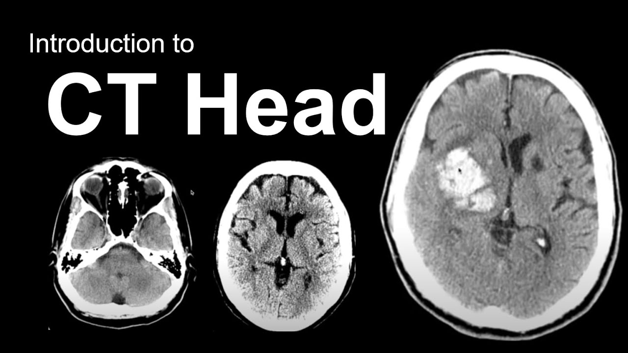
Introduction to CT Head: Approach and Principles

Abdominal Pain: Signs, Examination & Diagnosis – Emergency Medicine | Lecturio

Urinary/Kidney Stones - Overview (signs and symptoms, risk factors, pathophysiology, treatment)

Inguinal hernia anatomy

Colon Cancer (CRC) Signs & Symptoms (& Why They Occur)
Organs of the Human Body Songs | Anatomy Education Songs
5.0 / 5 (0 votes)
Thanks for rating: