Urinary/Kidney Stones - Overview (signs and symptoms, risk factors, pathophysiology, treatment)
TLDRThe video script delves into the subject of kidney stones, also known as urinary stones, which can form in various parts of the urinary tract. It explains the anatomy of the urinary system, including the kidneys, ureters, and bladder, and how kidney stones can form due to the accumulation of electrolytes like calcium, uric acid, and oxalate. The script outlines the medical conditions and symptoms associated with kidney stones, such as renal colic and hematuria, and discusses the risk factors, including diet and dehydration. Diagnostic methods like urinalysis, X-rays, ultrasounds, and CT scans are mentioned, along with various treatment options ranging from conservative management for small stones to surgical interventions like percutaneous nephrolithotomy and extracorporeal shock wave lithotripsy for larger stones. The summary emphasizes the importance of understanding kidney stones for both prevention and treatment.
Takeaways
- 📚 Kidney stones, also known as urinary stones, are mineral and acid deposits that can form in the urinary tract or kidneys.
- 🔍 The urinary tract includes the kidneys, ureters, bladder, and urethra, with specific sites of constriction where stones can lodge.
- 🧘♂️ Kidneys are vital organs that filter blood, regulate blood pressure, and maintain electrolyte balance, also producing important hormones.
- 🏠 The kidney's functional units, nephrons, are where blood filtration occurs, and the accumulation of electrolytes can lead to stone formation.
- 💠 If small, kidney stones may pass naturally in urine, but larger stones can cause obstructions, leading to severe pain known as renal colic.
- 🚨 Symptoms of kidney stones include acute flank pain, which may radiate to the back or groin, fever, nausea, vomiting, and hematuria.
- 🍽️ Risk factors for kidney stones include a high-protein or high-salt diet, obesity, dehydration, certain medications, and family history.
- 🧬 The formation of kidney stones is influenced by an increase in urinary solutes and a decrease in stone-forming inhibitors, leading to urine supersaturation.
- 📈 Diagnosis may involve blood tests, urinalysis, and imaging studies like X-rays, ultrasounds, or CT scans to detect and identify the type of stone.
- 🩺 Treatment options range from pain management for small stones that may pass naturally, to surgical interventions for larger stones causing obstructions.
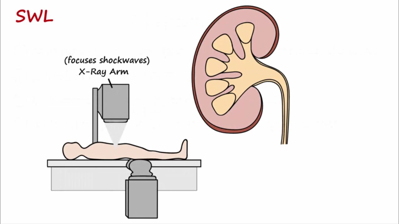
- ⚕️ Surgical management includes procedures like percutaneous nephrostomy, ureteral stent insertion, percutaneous nephrolithotomy, and extracorporeal shock wave lithotripsy.
Q & A
What are the different names for kidney stones?
-Kidney stones are also known as urinary stones, nephrolithiasis (stones forming in the nephron or kidneys), and renal or urinary calculi.
What are the three sites of constriction in the ureter where stones can lodge?
-The three sites of ureteral constriction include the pelvic ureteric junction, the pelvic brim, and the vesicular ureteric junction.
What is the role of the kidneys in the body?
-The kidneys are responsible for filtering the blood, disposing of waste, regulating blood pressure, and regulating electrolyte balance. They also produce important hormones such as erythropoietin and activate vitamin D.
What are the components of the kidney's functional unit, the nephron?
-The nephron consists of the Bowman's capsule, the glomerulus, the proximal convoluted tubules, the loop of Henle, the distal convoluted tubules, and the collecting duct.
How do kidney stones form?
-Kidney stones form when crystal-like structures, which are precipitates of accumulated electrolytes, grow larger in the nephron instead of passing through the urine.
What is renal colic?
-Renal colic is a severe, stabbing pain caused by the obstruction and buildup of pressure within the tubules of the kidney, often due to a kidney stone.
What are the common clinical presentations of kidney stones?
-Common clinical presentations include acute flank pain that may radiate to the back or groin, fever, nausea, vomiting, urinary frequency, urgency, and hematuria.
What are the risk factors for developing kidney stones?
-Risk factors include a high-protein or high-salt diet, obesity, dehydration, certain medications, and family history. These factors can lead to increased urinary solute concentration and decreased stone-forming inhibitors, causing urine supersaturation.
What are the five broad types of kidney stones?
-The five types are calcium oxalate stones, calcium phosphate stones, struvite stones (common in chronic urinary tract infections), uric acid stones, and cysteine stones.
What diagnostic tests are used to detect kidney stones?
-Diagnostic tests include a full blood count, CRP, urinalysis, 24-hour urine analysis for calcium, phosphate, oxalate, urate, cysteine, and xanthine levels, X-ray, ultrasound, and CT scan.
What are the surgical management options for kidney stones?
-Surgical management options include percutaneous nephrostomy (for drainage), ureteral stent insertion (to bypass blockage), percutaneous nephrolithotomy (for stones >2 cm), endoscopic procedures to break down stones, open surgery, and extracorporeal shock wave lithotripsy.
What is extracorporeal shock wave lithotripsy and how does it work?
-Extracorporeal shock wave lithotripsy (ESWL) is a non-invasive procedure that uses shock waves to break up stones in the kidneys, allowing the fragments to be passed out of the body through urine.
Outlines
😀 Understanding Kidney Stones and Their Anatomy
This paragraph introduces kidney stones, also known as urinary stones, and their various names such as Nephrolithiasis and urolithiasis. It explains the anatomy related to kidney stones, including the adrenal glands, kidneys, ureters, bladder, and urethra. The paragraph also describes the three sites of ureter constriction where stones can lodge. The kidneys' role in filtering blood, regulating blood pressure, and producing hormones is highlighted. The structure of the kidney, including the medulla, cortex, calyx, and renal pelvis, is detailed. Nephrons, the functional units of the kidney that filter blood and regulate electrolyte and fluid balance, are also explained. The paragraph concludes by discussing how crystals can form in the nephrons, potentially leading to urinary stones if they grow large enough to not pass through the urine.
😖 Symptoms and Risk Factors of Kidney Stones
The second paragraph delves into the symptoms and risk factors associated with kidney stones. It describes how kidney stones can cause obstruction and the resulting buildup of pressure, leading to renal colic and inflammation. The paragraph outlines the clinical presentation of kidney stones, which includes acute flank pain, fever, nausea, vomiting, urinary frequency, urgency, and hematuria. Risk factors for developing kidney stones are also discussed, such as a high-protein diet, high salt intake, obesity, dehydration, certain medications, and family history. The paragraph explains how these risk factors can lead to increased urinary solute concentration and decreased stone-forming inhibitors, resulting in urine supersaturation and crystal formation. It also categorizes kidney stones into five types: calcium oxalate, calcium phosphate, struvite, uric acid, and cysteine stones. Lastly, the paragraph mentions investigations for suspected renal calculi, including blood tests, urinalysis, and measurement of 24-hour urine levels of various substances.
🛠️ Diagnosis and Treatment of Kidney Stones
The final paragraph focuses on the diagnosis and treatment options for kidney stones. It lists diagnostic methods such as X-rays, ultrasounds, and CT scans to detect kidney stones and identify conditions like hydronephrosis. The paragraph describes the clinical presentation of kidney stones, emphasizing the acute flank pain and the triad of fever, vomiting, and acute flank pain. It also discusses the initial treatment approach, which includes analgesia, antiemetics, and IV fluids. The paragraph outlines various surgical interventions for kidney stones, such as percutaneous nephrostomy for drainage, ureteral stent insertion to bypass blockages, percutaneous nephrolithotomy for larger stones, endoscopic procedures to break down stones, open surgery, and extracorporeal shock wave lithotripsy to fragment stones for easier passage. The paragraph concludes by noting that most small urinary stones will pass spontaneously without intervention.
Mindmap
Keywords
💡Kidney stones
💡Ureter
💡Nephron
💡Renal colic
💡Urine supersaturation
💡Calcium oxalate stones
💡Urinary tract infection (UTI)
💡Hematuria
💡Percutaneous nephrolithotomy
💡Ureteroscopy
💡Extracorporeal shock wave lithotripsy (ESWL)
Highlights
Kidney stones, also known as urinary stones, have various names including nephrolithiasis and can form in the urinary tract or specifically in the kidneys.
The urinary tract includes the kidneys, ureters, bladder, and urethra, with three sites of constriction where stones can lodge.
The kidneys are vital for filtering blood, regulating blood pressure, and electrolyte balance, and also produce important hormones.
The nephron, the functional unit of the kidney, is responsible for filtering blood and reabsorbing necessary substances.
Urinary stones form when crystal-like structures, precipitates of accumulated electrolytes, grow larger in the nephron.
Kidney stones can cause renal colic, an obstruction, and inflammation, leading to severe pain that can radiate to the back or groin.
Risk factors for kidney stones include a high-protein diet, high salt intake, obesity, dehydration, and certain medications.
Urine supersaturation with stone-forming salts, such as calcium, uric acid, and oxalate, leads to crystal formation and potentially kidney stones.
Kidney stones can be categorized into five main types: calcium oxalate, calcium phosphate, struvite, uric acid, and cysteine stones.
Diagnostic methods for kidney stones include blood tests, urinalysis, 24-hour urine tests, X-ray, ultrasound, and CT scan.
Small urinary stones may pass spontaneously, while larger ones may require surgical intervention.
Surgical management options include percutaneous nephrostomy, ureteral stent insertion, percutaneous nephrolithotomy, endoscopic procedures, and open surgery.
Extracorporeal shock wave lithotripsy is a non-invasive procedure that uses shock waves to break up kidney stones.
The clinical presentation of kidney stones can include acute flank pain, fever, nausea, vomiting, hematuria, and urinary frequency.
In acute settings, treatment typically involves analgesia, antiemetics, and IV fluids, with careful monitoring.
Dehydration and changes in urinary pH contribute to urine supersaturation, increasing the risk of kidney stone formation.
Family history and certain medications can also increase the risk of developing kidney stones.
The anatomy of the urinary tract and its function in filtering and waste disposal is crucial to understanding kidney stone formation.
Transcripts


5.0 / 5 (0 votes)
Thanks for rating: