Unit 21: Acoustic Artifacts
TLDRThis educational video script delves into the world of ultrasound imaging, explaining what acoustic artifacts are and how they can distort true anatomy in images. It covers the common causes of artifacts, such as the machine's assumptions about sound travel and tissue interaction, and the impact of mechanical and operator errors. The script categorizes artifacts into resolution, position, and attenuation types, detailing each with examples and potential corrections. It also touches on the importance of recognizing and understanding artifacts for accurate clinical imaging.
Takeaways
- 🌐 An ultrasound artifact is any image feature that doesn't represent true anatomy, often due to the machine's assumptions about sound behavior in tissues being incorrect.
- 🔍 The machine assumes a constant sound speed of 1540 meters per second in soft tissues, but variations in this speed can lead to artifacts like misplacement of anatomical features.
- 🛠️ Mechanical and operator errors, such as a broken crystal or excessive gain reduction, can also cause artifacts by distorting or obscuring true anatomy.
- 🔎 There are different types of artifacts including resolution, position, and attenuation artifacts, each affecting the image in specific ways like blurring or misplacement.
- 📐 Resolution artifacts are related to the clarity and detail of the image, affected by factors like beam width and pulse length, resulting in issues like point spread or slice thickness artifacts.
- 📍 Position artifacts occur when anatomy appears in the wrong location, often due to sound changing direction (e.g., refraction, mirror artifact, multi-path artifact, reverberation, and ring down).
- 🔻 Attenuation artifacts happen when sound is absorbed or scattered by tissues to varying degrees, leading to shadowing, edge shadow, enhancement, and speckle.
- 👁️ Lateral resolution is crucial for distinguishing separate objects side by side and is affected by the width of the sound beam, impacting the clarity of the image.
- 🔄 To reduce artifacts, sonographers can adjust the transducer angle, frequency, depth, and gain, as well as utilize machine features like spatial compounding and frequency compounding.
- 📚 Understanding the physical principles behind artifacts helps in recognizing them and deciding whether to correct them or use them for diagnostic purposes.
- 🛑 Electronic and biological interference from external sources can also cause artifacts, which can sometimes be mitigated by using different frequency transducers or adjusting the imaging settings.
Q & A
What is an artifact in the context of ultrasound imaging?
-An artifact in ultrasound imaging is anything in the image that does not represent the true anatomy. It can cause fake anatomy to appear, real anatomy to disappear, or distort the shape, size, location, or brightness of the actual anatomy.
Why do artifacts occur in ultrasound imaging?
-Artifacts occur due to the machine making assumptions about how sound interacts with tissues, which can become invalid under certain conditions. This includes assumptions about the speed of sound, the direction of sound travel, and the nature of reflections, among others.
What is the typical assumption made by the ultrasound machine regarding the speed of sound in soft tissue?
-The typical assumption made by the ultrasound machine is that sound travels at 1540 meters per second in soft tissue, which is considered the propagation speed for soft tissue.
What are the different types of artifacts based on their effects on the image or anatomy?
-Artifacts can be categorized into resolution artifacts, position artifacts, and attenuation artifacts based on their effects on the image or anatomy.
How can poor axial resolution lead to an artifact in ultrasound imaging?
-Poor axial resolution can lead to an artifact when two objects that are parallel to the sound beam and sit less than half the spatial pulse length from one another are displayed as one object, not accurately representing the true anatomy.
What is a slice thickness artifact and how does it occur?
-A slice thickness artifact occurs when the ultrasound beam is too thick, causing echo information from above or below the slice to be detected in the image. This results in a partial volume artifact or suction thickness artifact, where anechoic structures may show unexpected echoes due to the thickness of the slice.
What is a mirror artifact and how does it occur?
-A mirror artifact occurs when sound interacts with a strong specular reflector at an oblique incidence, causing the beam to be reflected towards another structure and then back to the transducer. The ultrasound machine interprets these reflections as coming from the main beam path, creating a deep, exact replica of the anatomy.
How can mechanical errors and operator errors contribute to artifacts in ultrasound imaging?
-Mechanical errors such as a broken crystal can cause dropouts, which do not represent true anatomy and are considered artifacts. Operator errors, such as decreasing the gain too much and causing anatomy to disappear, also result in artifacts.
What is a reverberation artifact and how does it appear in an ultrasound image?
-A reverberation artifact occurs when sound bounces between two reflectors, with each bounce redirecting some sound energy to the other reflector and some back to the transducer. This results in equally spaced, parallel reflectors appearing deeper into the image, resembling a step ladder.
What is the purpose of speckle in ultrasound imaging and how can it be reduced?
-Speckle in ultrasound imaging represents the tissue texture and is created by the interference of echoes. It adds a grainy appearance to the image. Speckle can be reduced by using a higher frequency transducer or by utilizing machine techniques such as spatial compounding, frequency compounding, or coded excitation.
Outlines
🌐 Understanding Acoustic Artifacts in Ultrasound
This paragraph introduces the concept of acoustic artifacts in ultrasound imaging, explaining them as any image element that does not accurately represent true anatomy. Artifacts can either create false anatomy or cause the disappearance of real anatomy, often due to the machine making assumptions about sound travel that are invalidated by actual tissue interactions. The assumption of a constant sound speed of 1540 meters per second in soft tissue is highlighted, along with other assumptions that can lead to artifacts, such as direct sound travel to reflectors, unidirectional reflections, fixed sound direction, and a narrowly defined beam. The paragraph also mentions mechanical and operator errors as sources of artifacts and categorizes them into resolution, position, and attenuation artifacts.
🔍 Resolution Artifacts and Their Impact on Image Accuracy
The second paragraph delves into resolution artifacts, which occur when image details fail to accurately represent true anatomy due to the beam's insufficient narrowness in various planes. It discusses axial, lateral, and elevational resolutions, explaining how they relate to the spatial pulse length and beam width. The summary highlights the consequences of poor resolution, such as the merging of separate objects into one in the image, and provides strategies to mitigate these artifacts, such as using higher frequency transducers and positioning the focal point correctly. The paragraph also reviews the physics behind resolution artifacts and emphasizes the importance of understanding and correcting them for clinical imaging.
📍 Position Artifacts: Misplaced Anatomy in Ultrasound
This paragraph examines position artifacts, which cause anatomy to appear in incorrect locations due to sound direction changes. It covers various types of position artifacts, including refraction, mirror artifact, multi-path artifact, and others, that result from sound interacting with structures at oblique angles or reflecting off strong specular reflectors. The summary explains how these artifacts can create false replicas of anatomy and discusses the importance of the machine's timing mechanism in mapping echo returns to their paths. Strategies to resolve some of these artifacts, such as changing transducer position or understanding acoustic physics, are also mentioned.
🔄 Multi-Path and Reverberation Artifacts: Sound Redirection in Ultrasound
The fourth paragraph focuses on multi-path and reverberation artifacts, which occur when sound changes direction before returning to the transducer. It differentiates between these artifacts based on the reflectors involved and the resulting image patterns. The summary explains that multi-path artifacts create similar but not exact replicas of anatomy deep to the true structure, while reverberation artifacts produce equally spaced reflectors due to sound bouncing between two strong reflectors. The paragraph also discusses methods to mitigate these artifacts, such as adjusting the angle of insonation and reducing gain.
🌌 Reverberation and Ring Down Artifacts: Sound Interaction with Reflectors
This paragraph explores reverberation and ring down artifacts, which are caused by sound bouncing between reflectors. Reverberation artifacts are characterized by equally spaced lines parallel to the sound beam, while ring down artifacts appear as dense lines due to sound interacting with very small structures. The summary describes how these artifacts can aid in diagnosing certain conditions, such as the presence of air, and discusses the importance of understanding the physics behind these artifacts to interpret images accurately.
📉 Speed Error and Range Ambiguity Artifacts: Impact of Sound Propagation Speed
The sixth paragraph discusses speed error and range ambiguity artifacts, which are related to the speed at which sound travels through different mediums. Speed error artifacts occur when the sound speed deviates from the assumed 1540 meters per second, causing structures to appear shallower or deeper than they are. Range ambiguity artifacts happen when echoes return after a new pulse is sent, causing them to appear in the wrong location. The summary explains how these artifacts can affect image interpretation and suggests adjusting imaging depth and using different transducers to mitigate them.
🌑 Attenuation Artifacts: Effects of Sound Energy Reduction
This paragraph examines attenuation artifacts, which occur when sound energy is greatly or minimally affected by a reflector. It describes how shadowing, edge shadow, and enhancement artifacts are formed due to the interaction of sound with highly attenuating or low attenuation structures. The summary highlights the diagnostic value of these artifacts in identifying calcified structures and fluid-filled pathologies and discusses strategies for working around them to obtain clearer images.
🔆 Enhancement and Speckle Artifacts: Reflectivity and Texture Representation
The eighth paragraph discusses enhancement artifacts, which occur when sound energy is minimally attenuated, resulting in brighter echoes behind certain structures, and speckle, which is a grainy appearance on the image due to interference and represents tissue texture. The summary explains how enhancement can help distinguish fluid-filled structures from solid pathologies and how speckle can be reduced using higher frequency transducers for clearer images.
🛠 Techniques to Reduce Artifacts in Ultrasound Imaging
The final paragraph summarizes various techniques to reduce artifacts in ultrasound imaging, including spatial compounding, frequency compounding, and coded excitation. It emphasizes the importance of adjusting imaging settings and using different frequencies to minimize interference and speckle. The summary also encourages practitioners to continually assess and adjust their imaging techniques to ensure the highest image quality and artifact recognition.
Mindmap
Keywords
💡Acoustic Artifacts
💡Assumptions of Ultrasound Machine
💡Resolution Artifacts
💡Position Artifacts
💡Attenuation Artifacts
💡Mechanical and Operator Errors
💡Refraction
💡Mirror Artifact
💡Reverberation Artifact
💡Speckle
Highlights
An ultrasound artifact is any image detail that doesn't represent true anatomy, often caused by the machine's assumptions about sound interaction with tissues.
The assumption of sound traveling at 1540 meters per second in soft tissue can lead to artifacts if the actual speed varies.
Machines assume sound travels directly, doesn't change direction, and the beam is narrow, which can result in artifacts if these assumptions are invalid.
Mechanical and operator errors, such as a broken crystal or gain adjustment, can cause artifacts like dropout or anatomy disappearance.
Artifacts can be categorized into resolution, position, and attenuation artifacts based on the underlying assumptions and their effects on the image.
Resolution artifacts occur due to the beam not being narrow enough in all planes, affecting axial, lateral, and elevational resolution.
Lateral resolution can be improved by placing the area of interest within the focal zone of the beam for better image accuracy.
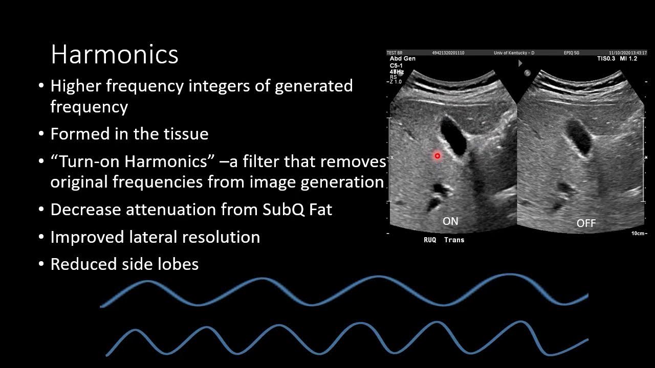
Elevational resolution issues manifest as slice thickness artifacts, which can be corrected using specific transducers or harmonics.
Position artifacts occur when anatomy appears in the wrong location due to sound changing directions or not returning directly to the transducer.
Refraction artifacts create exact replicas of anatomy placed laterally, caused by sound bending upon entering the body.
Mirror and multi-path artifacts occur when sound interacts with strong specular reflectors at oblique angles, creating deep replicas.
Reverberation artifacts are seen as equally spaced reflectors deep in the image, caused by sound bouncing between two strong reflectors.
Ring down or comet tail artifacts are dense lines appearing due to sound bouncing between very small structures.
Lobe artifacts display echoes from lateral anatomy as if they were in the main beam, affecting structures like the gallbladder or heart chambers.
Speed error artifacts are caused by the medium's propagation speed differing from the assumed 1540 meters per second, affecting echo placement.
Range ambiguity artifacts occur when echoes return after the next pulse is sent, causing them to appear in the wrong frame.
Attenuation artifacts, such as shadowing, edge shadow, and enhancement, result from sound energy being greatly or minimally affected by reflectors.
Speckle, the grainy appearance in ultrasound images, is technically an artifact representing tissue texture but can be reduced for clearer images.
Modern ultrasound machines have techniques like spatial compounding and frequency compounding to reduce artifacts and improve image quality.
Electronic and biological interference from external sources can cause artifacts in ultrasound images.
Sonographers should adjust settings like gain, depth, and focus to optimize image quality and minimize artifacts.
Transcripts
Browse More Related Video
Ultrasound Artifacts | Ultrasound Physics Course | Radiology Physics Course #25

Ultrasound Physics Review | What Are Artifacts | Sonography Minutes

Ultrasound Physics Review | Doppler Artifacts | Sonography Minutes

Ultrasound Physics Review | Velocity Error Artifacts | Sonography Minutes

Ultrasound Physics Review | Beam Artifacts | Sonography Minutes
Ultrasound Physics - Image Generation
5.0 / 5 (0 votes)
Thanks for rating: