Big Guns: The Muscular System - CrashCourse Biology #31
TLDRThis video explores how our muscles work on a cellular level. It examines the anatomy of skeletal muscles, including muscle fibers, myofibrils, sarcomeres, and myofilaments like actin and myosin. It explains how these proteins slide across one another when triggered by an action potential, using ATP as an energy source. This sliding filament model contracts the sarcomere and ultimately the muscle. The video further details the roles of ions like calcium in muscle contraction, and how ATP is also needed to relax the muscle after contraction. Overall, it reveals the complex molecular dance underlying our muscle movements.
Takeaways
- 😊 There are 3 types of muscles: cardiac, smooth, and skeletal. Skeletal muscles allow movement by contracting and relaxing.
- 👉 Skeletal muscles are made up of layers: muscle fibers containing myofibrils with sarcomeres that contract.
- 🧠 Muscle contraction works via the 'sliding filament model' - actin and myosin filaments slide across one another.
- 🔋 ATP provides energy for muscle contraction. Myosin heads bind to actin, powered by ATP.
- 💪 When myosin binds actin, the sarcomere contracts, shortening the muscle.
- 🛑 For the muscle to relax, ATP attaches to myosin heads, breaking the actin bonds.
- ⚡ Action potentials trigger calcium ion release, initiating contraction.
- 📈 Concentration gradients allow calcium pumping/storage to reset the muscle.
- 📚 We didn't understand how muscles contract until research in 1954 established the sliding filament model.
- 👍 It's complex, but understanding muscle chemistry allows movement - from dance moves to lifting weights!
Q & A
What are the three different types of muscle in the human body?
-The three different types of muscle in the human body are: 1) Cardiac muscle, found in the heart, 2) Smooth muscle, responsible for involuntary processes like digestion and blood flow, and 3) Skeletal muscles, which allow for voluntary movement of the body.
What makes meat stringy in texture?
-The layered structure of skeletal muscle fibers is what makes meat stringy. Skeletal muscles are made up of layers of muscle fascicles, which are bundles of long, thin muscle fibers (muscle cells).
How many strands of actin are typically in a sarcomere?
-While the number varies by muscle type, the video states that in this particular example there are four actin strands in the sarcomere - two sitting on top and two sitting on the bottom.
What stops the myosin from gripping the actin when the muscle is at rest?
-When the muscle cell is at rest, tropomyosin and troponin proteins wrap around and cover the binding sites on the actin strands. These proteins act as 'chaperones' to prevent the myosin heads from binding to the actin.
What provides the stimulus for a muscle contraction to start?
-A muscle contraction begins when a signal travels down a motor neuron to the synapse with a muscle cell. This triggers neurotransmitters to be released, starting an action potential in the muscle cell.
How does calcium help lead to muscle contraction?
-The sarcoplasmic reticulum stores calcium ions when a muscle cell is at rest. When signaled, it releases these ions which bind to troponin and remove tropomyosin from the actin - allowing myosin to bind and muscle contraction to occur.
What makes a myosin head bind to and release actin?
-Myosin heads have pent up energy from a spent ATP molecule that they use to bind to actin. When a new passing ATP molecule attaches, its energy release breaks the bond and causes myosin to release the actin.
Why does rigor mortis happen?
-After death, there is no more ATP being produced so myosin heads stay tightly bound to actin. With calcium ions also leaking out to signal contraction, the muscles are stuck contracted.
How is energy from ATP used both for muscle contraction and relaxation?
-The pent up energy in used ATP attached to myosin allows myosin to bind to actin and cause contraction. New ATP energy is then needed to cause myosin to release actin again and allow muscle relaxation.
What two proteins must be removed or shift positions for muscle contraction to occur?
-Tropomyosin and troponin must shift position or be removed from binding sites on actin to allow myosin heads to grip actin and cause contraction.
Outlines
😄 Anatomy of Skeletal Muscles
The first paragraph introduces skeletal muscles - the type of muscles used for voluntary movement. It describes the structure of a skeletal muscle, including the muscle belly, tendons, muscle fascicles, muscle fibers, and sarcomeres. It explains how sarcomeres contract and relax to create muscle movement.
👩🔬 Sliding Filament Model of Muscle Contraction
The second paragraph explains the sliding filament model of muscle contraction proposed in 1954. It involves the thick myosin and thin actin protein filaments sliding over each other and the role of ATP in powering this process. Tropomyosin and troponin proteins regulate the interaction.
⚡ Signal Transduction in Muscle Contraction
The third paragraph details the series of events that transduce a neural signal to cause muscle contraction. It covers action potentials, calcium ion release, myosin binding to actin, power stroking, relaxation aided by ATP, and calcium ion pumping back into the sarcoplasmic reticulum.
Mindmap
Keywords
💡muscles
💡contraction
💡myosin
💡actin
💡ATP
💡sarcomere
💡calcium ions
💡motor neuron
💡rigor mortis
💡sliding filament model
Highlights
Muscles are only good at two things: contracting to become shorter, and relaxing back out to their resting length.
Muscle cells are basically just bundles of complex protein strands, and since nuclei are essential for the protein-making process, muscle cells need lots of nuclei.
The chemical dance that allows muscles to contract is one of the sexiest things that goes on in your body, other than, like, sex, and it's known as the sliding filament model of muscle contraction.
We didn't figure out how muscles worked until 1954 when two teams discovered that the sliding filament model is how muscles contract.
Muscle contractions are caused by the movement of myosin protein filaments over actin protein filaments inside muscle cells.
ATP creates the energy for almost everything your body does, including muscle movement.
When a nerve signal reaches the muscle cell, it triggers calcium ions to be released, allowing myosin heads to bind to actin and cause contraction.
It's the energy from ATP that actually makes the muscle relax after contraction.
Rigor mortis happens because without ATP, the muscles get stuck in the contracted state.
Myosin heads repeatedly bind to actin, get hit with an ATP molecule, release energy to relax, detach, and then start the process over.
Chemistry makes all muscle contraction and relaxation possible, from lifting weights to dance moves.
Muscles are connected to bones by tendons, while ligaments connect bones to other bones.
Each muscle cell has tens of thousands of sarcomeres that contract together when stimulated.
Troponin and tropomyosin act as "chaperones" that keep myosin from binding to actin when the muscle is at rest.
The sarcoplasmic reticulum stores calcium ions and releases them when a nerve signal comes to trigger contraction.
Transcripts
Browse More Related Video

Muscles, Part 1 - Muscle Cells: Crash Course Anatomy & Physiology #21
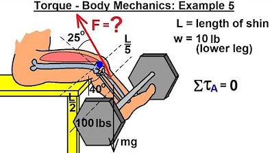
Physics 15 Torque (17 of 25) Body Mechanics: Ex. 5, F=? Leg Lifting Weights

Fermentation

Physics 15 Torque (14 of 27) Body Mechanics: Ex. 2, F=? To Lift Dumbbell

Fluid and Electrolytes for Nursing Students - Comprehensive NCLEX Review
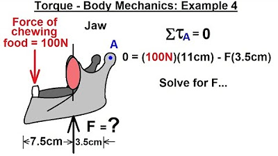
Physics 15 Torque (16 of 25) Body Mechanics: Ex. 4, F=? Jaw Muscle
5.0 / 5 (0 votes)
Thanks for rating: