ARDMS Ultrasound Physics
TLDRThis educational video script delves into ultrasound imaging techniques, focusing on how various factors impact image resolution and quality. It clarifies misconceptions about image storage post-digitization and the sequence of events in ultrasound imaging. The script also explores the creation of harmonics in deeper tissues, the effects of pulse inversion, and strategies to improve frame rate. Additionally, it provides insights into Doppler phantom quality assurance and the calculation of positive predictive value, offering valuable knowledge for those preparing for ultrasound board exams.
Takeaways
- 📉 Using pulse inversion in ultrasound imaging increases spatial resolution but decreases temporal resolution.
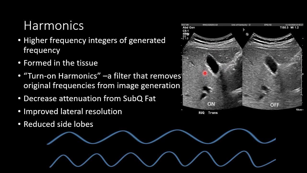
- 🌀 Harmonics are created in deeper tissue areas due to the non-linear behavior of sound waves traveling through tissues of varying density.
- 🔍 Fundamental imaging is a type of non-linear imaging that helps reduce artifacts by incorporating higher harmonics.
- 📊 B-mode is used to demonstrate the superposition of different imaging modes, such as B mode over A mode.
- 🚫 Pulse repetition frequency is not associated with adding an extra pulse per scan line, unlike other techniques like multi-focusing packets.
- 💾 After an image is digitized, it is first displayed on the screen before being stored in PACS.
- 🔑 Understanding the difference between 'what happens first after an image is digitized' and 'where is it stored after it's digitized' is crucial for answering related questions correctly.
- 📈 To improve frame rate, decreasing the depth of the image is suggested, as opposed to increasing line density or using other imaging techniques.
- 📊 The positive predicted value (PPV) in a Doppler phantom's quality assurance chart is calculated by dividing true positives by the sum of true positives and false positives.
- 🛑 M-mode is identified as a technique that does not degrade frame rate and offers good temporal resolution.
- ⏪ To fix attenuation artifacts, compression is recommended over other gain compensation techniques.
- 📏 The y-axis in B-mode does not represent time or brightness; instead, B-mode has an x-axis for lateral movement and a z-axis for depth.
Q & A
What degrades the temporal resolution when using harmonic imaging?
-The degradation of temporal resolution occurs when using pulse inversion. This technique adds an extra pulse to a scan line, which increases spatial resolution but decreases temporal resolution as a result.
What are the other methods that can add an extra pulse to a scan line similar to pulse inversion?
-Other methods that add an extra pulse to a scan line include multi-focusing packets and ensemble length packets, which are essentially the same thing.
Where are harmonics typically created in the body?
-Harmonics are typically created in the deeper tissue. They are not generated in the superficial region as the pulse travels from superficial to deep areas, encountering different densities and creating harmonics due to non-linear behavior.
What is the difference between fundamental imaging and non-linear imaging?
-Fundamental imaging involves the use of the original frequency without higher harmonics, while non-linear imaging includes higher harmonics and can result in more artifacts.
What imaging mode is represented by the acronym 'B mode'?
-B mode stands for brightness mode, which is a type of ultrasound imaging that displays the brightness of echoes on a two-dimensional screen.
What does the term 'PPV' stand for in the context of a Doppler phantom's quality assurance?
-PPV stands for Positive Predictive Value, which is calculated as the number of true positives divided by the sum of true positives and false positives.
What is the first thing that happens to an image after it is digitized?
-After an image is digitized, the first thing that happens is that it is displayed on the screen. It then goes to storage in PACS.
What will not degrade the frame rate according to the script?
-Using a smaller sector, M-mode, and compression will not degrade the frame rate. Continuous wave Doppler, 3D imaging, persistence, and load duty factor are more likely to degrade frame rate.
What does the Y-axis on B-mode represent?
-There is no Y-axis in B-mode. B-mode has an X-axis for lateral movement and a Z-axis for depth.
How can one improve frame rate in an ultrasound image?
-To improve frame rate in an ultrasound image, one can decrease the depth of the scan, as increasing line density or using color Doppler might degrade the frame rate.
What technique will not fix attenuation artifacts in ultrasound imaging?
-Compression will not fix attenuation artifacts. Techniques like time gain compensation or swept gain are more suited for addressing such artifacts.
Outlines
🌟 Ultrasound Imaging Techniques and Artifacts
The first paragraph introduces a quiz on ultrasound imaging, focusing on the effects of pulse inversion on spatial and temporal resolution. It explains how adding an extra pulse to a scan line can increase spatial resolution but decrease temporal resolution. The paragraph also discusses the creation of harmonics in deeper tissues due to non-linear behavior of sound waves and how fundamental imaging can reduce artifacts. It touches on different imaging modes, such as A-mode, B-mode, M-mode, and Z-mode, and their characteristics.
📈 Impact of Imaging Techniques on Frame Rate and Quality Assurance
This paragraph delves into the impact of various ultrasound imaging techniques on frame rate, highlighting that continuous wave Doppler does not degrade it, unlike other methods. It also addresses where images are stored after digitization, emphasizing the importance of understanding the sequence of events post-digitization. The paragraph further discusses improving frame rate by decreasing depth and introduces a quality assurance chart for Doppler phantoms, explaining the concept of positive predictive value (PPV) and how it is calculated.
📞 Ultrasound Board Exam Preparation and Resources
The final paragraph offers assistance for those preparing for their ultrasound board exams, providing contact information for further questions. It promotes additional study materials, mock exams, and tutoring services available through the website ultrasoundboardview.com. The speaker, Jim, encourages viewers to reach out for tips and advice to successfully pass their board exams and thanks them for watching the video.
Mindmap
Keywords
💡Harmonics Imaging
💡Pulse Inversion
💡Fundamental Imaging
💡Non-linear Behavior
💡B-Mode
💡M-Mode
💡Doppler Phantom
💡Positive Predictive Value (PPV)
💡Frame Rate
💡Persistence
💡SPI
Highlights
Introduction to a special ultrasound physics quiz session.
Degradation of temporal resolution with pulse inversion in harmonic imaging.
Explanation of how adding an extra pulse to a scan line increases spatial resolution but decreases temporal resolution.
Other techniques that add extra pulses to a scan line, such as multi-focusing and ensemble length packets.
Harmonics are created in deeper tissue, not in the superficial region.
The creation of harmonics is due to non-linear behavior of sound traveling through tissues of varying density.
Fundamental imaging is associated with non-linear imaging and reduced artifacts.
Identification of B-mode as the imaging mode demonstrated in the quiz.
Description of an image showing B-mode superimposed over A-mode.
Pulse repetition frequency is not associated with more than one pulse per scan line.
Continuous wave Doppler is identified as not degrading frame rate.
The process of an image being digitized and then displayed before being stored in PACS.
The importance of reading questions carefully to understand the sequence of events after image digitization.
Smaller sector size as a method to improve frame rate without degradation.
Explanation of the Positive Predictive Value (PPV) calculation in the context of a Doppler phantom quality assurance.
M-mode identified as a method that does not degrade frame rate and has good temporal resolution.
Compression as a technique that does not fix attenuation artifacts.
Clarification that the Y-axis in B-mode does not represent time or brightness but is absent.
Contact information provided for questions about upcoming SBI boards and additional study materials.
Promotion of mock exams and tutoring services for SPI preparation.
Closing remarks with an offer of tips and advice for successfully passing the boards.
Transcripts
Browse More Related Video

SPI Review

Tissue Harmonic Ultrasound Imaging | Ultrasound Physics Course | Radiology Physics Course #24

Ultrasound Physics Registry Review

Ultrasound Physics - Image Generation

Ultrasound Transducer (Part 2) Damping Block and Transducer Wiring | Ultrasound Physics #10

Ultrasound Physics Review | Resolution | Sonography Minutes
5.0 / 5 (0 votes)
Thanks for rating: