Lec-33 I Electron Microscopy I Applied chemistry I Chemical engineering
TLDRThe video lecture series on applied chemistry focuses on electron microscopy as an advanced analytical technique for compound identification at the nanoscale. It differentiates between scanning electron microscopy (SEM) and transmission electron microscopy (TEM), highlighting SEM's role in surface analysis and TEM's in internal structure examination. The lecture delves into the principles and components of SEM, its operation, and its detectors. The applications of SEM are vast, including semiconductor inspection, microchip production, and research in various scientific fields, emphasizing its significance in modern technology and research.
Takeaways
- 🧪 The video lecture series focuses on applied chemistry, with a specific subject goal of 3130506.
- 📚 In previous sessions, two analytical techniques were discussed: NMR spectroscopy and mass spectrometry, both sophisticated methods for compound identification.
- 🔬 This session introduces electron microscopy, an advanced technique for identifying compounds at the nanoscale level.
- 🔍 Electron microscopy uses a beam of high-energy electrons to examine objects at a fine scale, providing detailed information about the substance's topography, morphology, composition, and crystallography.
- 💡 The examination information provided by electron microscopes includes surface characteristics, structure of compounds, the presence of specific atoms or molecules, and detailed substance information.
- 📈 There are two main types of electron microscopes: Scanning Electron Microscopes (SEM) for surface visualization and Transmission Electron Microscopes (TEM) for studying internal structures.
- 🖱️ SEM requires a soundproof room, vacuum conditions, and high-definition computers to detect and process images.
- 🔧 Proper sample preparation and knowledgeable personnel are essential for effective scanning electron microscopy.
- 🎯 The scanning electron microscope operates by scanning a sample with a focused electron beam, producing secondary electrons that provide information about the sample's surface.
- 🔗 When SEM is combined with Energy-Dispersive X-ray Spectroscopy (EDS), it can determine both surface topography and composition.
- 🏭 SEM has extensive applications in various fields, including semiconductor inspection, microchip production, scientific research, gemology, medical and forensic sciences, and metallurgy.
Q & A
What is the subject goal for the video lecture series on applied chemistry?
-The subject goal for the video lecture series on applied chemistry is 3130506.
What are the two analytical techniques discussed in the previous sessions of chapter nine?
-The two analytical techniques discussed are Nuclear Magnetic Resonance (NMR) spectroscopy and Mass spectrometry.
What is the primary focus of electron microscopy in the context of the lecture?
-Electron microscopy focuses on the identification of compounds at very low levels, such as nanometers, by using a beam of electrons or highly energetic electrons to examine objects at a fine scale.
What kind of information can be obtained from electron microscopy?
-Electron microscopy can provide information about topography, morphology, composition, and crystallography of a substance.
What are the two main types of electron microscopes and what do they examine?
-The two main types are Scanning Electron Microscopes (SEM) which visualize the surface of objects, and Transmission Electron Microscopes (TEM) which study the internal structure of objects.
What are the key components and requirements for a scanning electron microscope (SEM) setup?
-Key components and requirements for SEM include a soundproof room, vacuum conditions, temperature control, vacuum pumps, supercomputers for high-definition imaging, a proper sample preparation kit, and a knowledgeable, trained person to handle the microscope.
How does a scanning electron microscope (SEM) produce images of a sample?
-SEM produces images by scanning the sample with a focused beam of electrons, which interact with the sample's electrons to produce secondary electrons that are then captured and used to identify the substance.
What is the role of the electron gun in SEM?
-The electron gun is responsible for producing high-speed, highly energetic electrons that are used to scan the sample and create an image.
What are the applications of scanning electron microscopy (SEM)?
-Applications of SEM include semiconductor inspection, production line analysis, microchip assembly, scientific research in biology and pharmaceuticals, gemology, medical and forensic sciences, and metallurgical operations.
How does SEM contribute to the study of microstructures and surface analysis?
-SEM can detect and analyze surface fractures, examine surface contaminations, reveal variations in chemical compositions, and provide qualitative and quantitative chemical analysis, which are crucial for understanding microstructures and surface properties.
What are the different detectors used in SEM and what do they capture?
-There are three main detectors in SEM: EDS (Energy-Dispersive X-ray Spectrometer), secondary electron detectors, and backscattered electron detectors. EDS determines surface composition, while the other two capture secondary and backscattered electrons for surface topography analysis.
Outlines
🔬 Introduction to Electron Microscopy Techniques
This paragraph introduces the topic of electron microscopy as a sophisticated method for compound identification at the nanoscale level. It contrasts electron microscopy with traditional optical microscopy and highlights the capabilities of electron microscopy in providing topography, morphology, composition, and crystallographic information about a substance. The paragraph also outlines the different types of electron microscopes, specifically scanning electron microscopes (SEM) and transmission electron microscopes (TEM), emphasizing their roles in surface and internal structure analysis respectively.
🏢 Requirements and Setup for Scanning Electron Microscopy (SEM)
This paragraph delves into the specific requirements and setup for using a scanning electron microscope. It emphasizes the need for a soundproof room with a vacuum environment and temperature control to maintain the integrity of the instrument. The paragraph also discusses the necessity of high-definition imaging capabilities, vacuum pumps, and proper sample preparation. Additionally, it underscores the importance of having knowledgeable personnel to operate the SEM and conduct compound identification. The paragraph explains how SEM works by scanning a sample with a focused beam of electrons, producing various signals that provide information about the sample's surface topography and composition.
🔧 Components and Functionality of Scanning Electron Microscope (SEM)
This paragraph describes the internal components and functionality of a scanning electron microscope. It outlines the roles of the electron gun, condenser lenses, objective aperture, scan coils, specimen stage, and various detectors. The paragraph explains how these components work together to produce high-resolution images of a sample's surface. It also details the process of electron interaction with the sample, the production of secondary and backscattered electrons, and their detection for image formation. The paragraph further discusses the different types of detectors used in SEM and their specific functions in capturing electrons for analysis.
📈 Practical Applications and Significance of Scanning Electron Microscopy (SEM)
This paragraph highlights the wide range of practical applications of scanning electron microscopy in various fields. It mentions its crucial role in industries such as semiconductor inspection, microchip production, and the analysis of nano-level structures. The paragraph also discusses SEM's importance as a research tool in scientific studies across disciplines like biology, pharmaceuticals, gemology, medical and forensic sciences, and metallurgy. It emphasizes the ability of SEM to detect surface fractures, examine contaminations, reveal chemical composition variations, and provide qualitative and quantitative chemical analysis, thus showcasing its significant impact on future technological and scientific advancements.
Mindmap
Keywords
💡Applied Chemistry
💡Analytical Techniques
💡Electron Microscopy
💡Scanning Electron Microscope (SEM)
💡Transmission Electron Microscope (TEM)
💡Surface Topography
💡Morphology
💡Composition
💡Electron Gun
💡Backscattered Electrons
💡Data System
Highlights
The subject goal for blood chemistry is 3130506.
In chapter nine, analytical techniques such as NMR spectroscopy and mass spectrometry were learned.
Electron microscopy is introduced as an advanced technique for compound identification at nano levels.
Electron microscopy provides information on topography, morphology, composition, and crystallography of substances.
Scanning Electron Microscope (SEM) and Transmission Electron Microscope (TEM) are the two types of electron microscopes.
SEM is used for surface visualization while TEM is for studying the internal structure of objects.
The setup for an electron microscope requires a soundproof, vacuum, and temperature-controlled environment.
High-definition imaging requires supercomputers and proper sample preparation for electron microscopy.
A knowledgeable, trained person is essential for handling electron microscopes.
Scanning Electron Microscopy (SEM) uses a focused beam of electrons to scan and produce images of the sample.
Incident electrons interact with the sample, producing secondary electrons that provide surface information.
The combination of SEM with EDS (Energy-Dispersive X-ray Spectrometer) allows for the determination of surface topography and composition.
Electron microscopy has a wide range of applications including semiconductor inspection and nanochip analysis.
SEM is essential in research tools for various fields such as biology, pharmaceuticals, gemology, medical and forensic sciences, and metallurgy.
SEM can detect and analyze surface fractures, contaminations, and chemical composition variations.
The internal structure of an electron microscope includes an electron gun, condenser lenses, objective aperture, scan coils, specimen stage, and computer hardware for data interpretation.
Different detectors are used in SEM to capture backscattered and secondary electrons for image formation.
Transcripts
Browse More Related Video
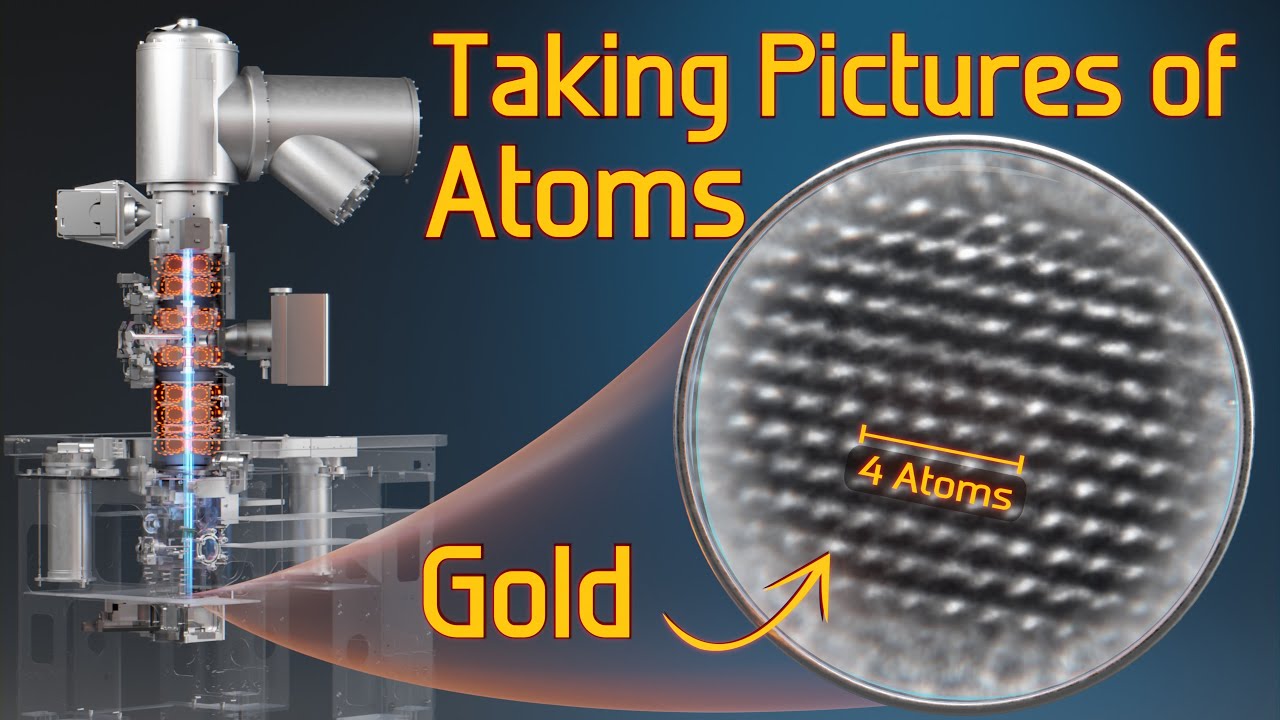
How do Electron Microscopes Work? 🔬🛠🔬 Taking Pictures of Atoms

Lec-32 l NMR Spectroscopy l Applied chemistry | Chemical engineering

19. Uses of Photon and Ion Nuclear Interactions — Characterization Techniques

50,000,000x Magnification

Lec-31 | Mass spectrometry | Applied chemistry | Chemical engineering

Applications (Contd.)
5.0 / 5 (0 votes)
Thanks for rating: