Human Eye - Passage of light through it | Don't Memorise
TLDRThis video delves into the journey of light within the human eye, explaining how we perceive our surroundings. Starting at the cornea, light is refracted and travels through the aqueous humor to the lens, which adjusts its shape for focus on the retina, forming an inverted image that the brain rights. The process involves multiple refractions, primarily at the air-cornea interface, and highlights the lens's accommodation ability to adjust focal length for clear vision at varying distances. The video concludes by introducing common eye defects to be discussed in the next installment.
Takeaways
- 👁 The human eye's simplified 2D model helps us understand how light propagates and allows us to see objects around us.
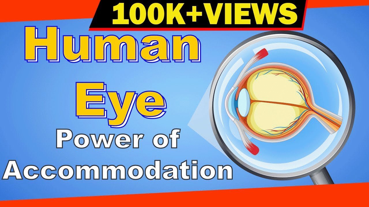
- 🌟 Light from an object first enters the eye and is refracted by the cornea, the transparent outer layer guarding the eye's entrance.
- 💧 The refracted light travels through the aqueous humor, a transparent fluid, before entering the pupil and reaching the lens.
- 🔍 The eye's lens, a biconvex lens, focuses the light so that it forms a real and inverted image on the retina.
- 🧠 Our brain corrects the inverted image, allowing us to perceive the world as it is, not upside down.
- 👀 Light signals are transmitted as impulses through the optic nerve to the occipital lobe of the brain.
- 🌌 The space between the lens and the retina is filled with vitreous humor, another transparent fluid.
- 🔬 Multiple refractions occur as light travels through different mediums, with the most significant refraction happening at the air-cornea interface.
- 🔧 The lens's shape can change, being controlled by ciliary muscles, to ensure light is focused on the retina, a process known as accommodation.
- 🔍 The lens adjusts its focal length by changing its thickness, allowing it to focus light on the retina for objects at varying distances.
- 🚫 The eye has a minimum distance for clear vision, approximately twenty-five centimeters, beyond which objects appear distorted if too close.
Q & A
What is the primary function of the cornea in the process of vision?
-The cornea is the curved and transparent outer layer that guards the entrance to the eye. Its primary function is to refract the light that enters the eye from an external object.
What is the role of the aqueous humor in the eye?
-The aqueous humor is a transparent fluid that fills the space between the cornea and the lens. It helps in the transmission of light from the cornea to the lens.
How does the lens of the eye contribute to the formation of an image on the retina?
-The lens, being a biconvex lens, refracts the light in such a way that all light coming from a point on an object is focused on the retina, forming a real and inverted image.
Why do we not perceive the world as being upside down, even though the image on the retina is inverted?
-The brain interprets the inverted image and processes it to appear upright, allowing us to perceive the world as it is.
What is the occipital lobe and how is it related to vision?
-The occipital lobe is a part of the brain where the optic nerve sends light signals as impulses. It is responsible for processing visual information.
What is vitreous humor and where is it located in the eye?
-Vitreous humor is a transparent gel-like substance that fills the space between the lens and the retina. It helps in maintaining the shape of the eye and in the transmission of light to the retina.
What are the ciliary muscles and how do they affect the eye's lens?
-The ciliary muscles are muscles that control the shape of the eye's lens. When they contract, the lens becomes thicker and its focal length decreases, allowing the eye to focus on nearby objects.
What is accommodation and how does it work in the human eye?
-Accommodation is the ability of the eye's lens to adjust its focal length to focus on objects at varying distances. It is achieved by the contraction and relaxation of the ciliary muscles, which change the lens's shape.
What happens when an object is too close to the eye, beyond the normal eye's minimum distance?
-When an object is too close, the image does not form on the retina, resulting in a distorted and unclear view. The eye may strain to reduce the focal length of the lens, but the object will still not be seen clearly.
What is the minimum distance of the object from the eye for a normal eye, and why is it significant?
-The minimum distance for a normal eye is around twenty-five centimeters. It is the closest distance at which an object can be placed without causing the image to be formed in front of the retina, ensuring clear vision.
What is the significance of the refractive indices of the different media through which light travels in the eye?
-The refractive indices of the media (air, cornea, aqueous humor, lens, and vitreous humor) determine the degree of refraction at each interface. The largest difference in refractive indices occurs at the air-cornea interface, which is where most of the refraction happens.
Outlines
👀 Understanding Light Propagation in the Eye
This paragraph explains the journey of light inside the human eye, starting with the cornea and moving through the aqueous humor to the lens. The lens's role in focusing light onto the retina to form an inverted image is highlighted. The brain's ability to correct this inversion and present a right-side-up image is also discussed. The paragraph delves into the refractive properties of the eye's media, emphasizing the significant refraction at the air-cornea interface. It further explains the lens's flexibility, controlled by the ciliary muscles, which allows for accommodation, ensuring that images are always focused on the retina regardless of the object's distance.
🔍 Addressing Eye Strain and the Concept of Minimum Distance
The second paragraph addresses the issue of eye strain when trying to see objects that are too close, which can distort the image and make it unclear. It explains that the eye's natural accommodation has limits and suggests maintaining an optimal distance from the eye to ensure clear vision. The 'minimum distance' concept is introduced, which is the ideal distance at which an object should be placed to form a clear image on the retina. The normal eye's minimum distance is approximately twenty-five centimeters. The paragraph concludes by summarizing the key points about light propagation in the eye and sets the stage for the next video, which will explore eye defects.
Mindmap
Keywords
💡Cornea
💡Refraction
💡Aqueous Humor
💡Pupil
💡Lens
💡Retina
💡Occlusion
💡Optic Nerve
💡Vitreous Humor
💡Accommodation
💡Ciliary Muscles
💡Minimum Distance
Highlights
The video explains the propagation of light within the human eye and the process of vision.
A simplified 2D model of the eye is used to illustrate how light enters and is processed.
Light first strikes the cornea, the transparent outer layer that guards the eye's entrance.
The cornea's refraction of light is the initial step in the process of vision.
Aqueous humor, the transparent fluid, directs light from the cornea to the pupil.
The biconvex lens of the eye focuses light onto the retina, forming a real and inverted image.
The brain corrects the inversion, allowing us to perceive the world as upright.
The optic nerve transmits light signals as impulses to the occipital lobe of the brain.
Vitreous humor, another transparent fluid, is located between the lens and the retina.
Multiple refractions occur within the eye, with the majority happening at the air-cornea interface.
The refractive indices of the media through which light travels are detailed, emphasizing the significance of the air-cornea difference.
The lens's primary role is to fine-tune light to ensure it focuses solely on the retina.
The lens's flexibility allows it to change shape and adjust focal length as needed.
Ciliary muscles control the lens's movement, affecting its thickness and focal length.
Accommodation is the lens's ability to adjust its focal length for different viewing distances.
There are limits to the lens's accommodation, affecting the clarity of nearby objects.
Straining the eyes attempts to reduce the lens's focal length but can distort vision.
The minimum distance for clear vision is the distance at which the image is optimally formed on the retina.
The video concludes by summarizing the light propagation process within the eye and the role of the lens and vitreous humor.
Transcripts
Browse More Related Video

Why Can't You Focus Underwater? | Physics with Professor Matt Anderson | M28-05
Power of accommodation of the Human Eye | Letstute

Eye defects - Myopia | Don't Memorise

Physics - Optics: Vision Correction (1 of 5) Introduction

Optical Instruments: Crash Course Physics #41

Some Of You Can See The Invisible
5.0 / 5 (0 votes)
Thanks for rating: