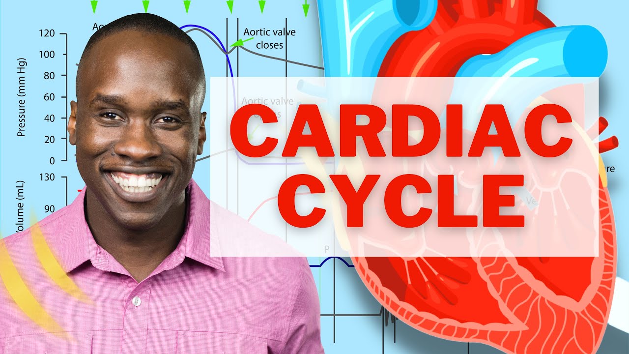
Cardiovascular | Cardiac Cycle
TLDRThe video script offers an in-depth exploration of the cardiac cycle, detailing the mechanical events that occur as blood flows through the heart's chambers. It emphasizes the cycle's duration of approximately 0.8 seconds and breaks down the process into key stages, including ventricular diastole, atrial and ventricular pressure differences, the role of atrioventricular (AV) valves and semilunar valves, and their impact on the EKG. The script explains how blood flow is regulated by pressure gradients across these valves, leading to the heart's characteristic sounds and the EKG's P wave, QRS complex, and T wave. The narrative concludes by illustrating the continuous nature of the cardiac cycle, highlighting its importance for distributing blood throughout the body.
Takeaways
- 🕒 The cardiac cycle refers to the series of mechanical events that occur as blood flows through the heart's chambers, taking about 0.8 seconds on average.
- 📉 Diastole is the phase of relaxation when the heart is filling with blood from the veins, and it's the period when the atria pressure exceeds the ventricles, leading to the opening of the atrioventricular (AV) valves.
- 🌀 During mid to late ventricular diastole, the SA node fires, causing the atria to depolarize and contract, pushing the remaining 20% of blood into the ventricles, which is represented by the P wave on the EKG.
- 🔄 Isovolumetric contraction is the phase where the ventricles start to contract but no blood is ejected yet; this phase is characterized by the closure of the AV valves and the production of the first heart sound, 'Lub' or S1.
- ➡️ Ventricular ejection occurs when the pressure in the ventricles exceeds the pressure in the arteries, causing the semilunar valves to open and blood to be ejected into the pulmonary trunk and aorta, represented by the QRS complex on the EKG.
- 🛑 At the end of ventricular systole, the ventricles have ejected most of their blood, and the remaining volume is known as the end systolic volume (ESV). The pressure in the arteries then exceeds the ventricular pressure, causing the semilunar valves to close and producing the second heart sound, 'Dub' or S2.
- 🔽 Isovolumetric relaxation marks the beginning of ventricular relaxation, where all valves are briefly closed, and the ventricles begin to refill in preparation for the next cardiac cycle.
- 🔄 The cardiac cycle repeats continuously, with the ventricles repolarizing and the T wave appearing on the EKG, indicating the end of the cycle and the beginning of the next.
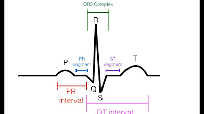
- 📝 The EKG is a crucial tool for visualizing the electrical activity of the heart, with different components corresponding to various stages of the cardiac cycle, such as the P wave for atrial depolarization and the T wave for ventricular repolarization.
- 🩸 Blood flow through the heart is a result of pressure differences, with blood naturally moving from areas of high pressure to areas of low pressure, regulated by the opening and closing of heart valves.
- 🧘🏼♂️ The heart's muscular layer, the myocardium, plays a vital role in the cardiac cycle by contracting to move blood through the heart and into the arteries.
Q & A
What is the cardiac cycle?
-The cardiac cycle refers to the sequence of mechanical events that occur as blood flows through the heart's chambers. It takes approximately 0.8 seconds on average to complete one cycle.
What is the difference between atrial and ventricular pressure during the cardiac cycle?
-Atrial pressure is slightly higher than ventricular pressure, which allows blood to flow from the atria into the ventricles when the atrioventricular (AV) valves open. Conversely, during ventricular contraction, ventricular pressure exceeds atrial pressure, causing the AV valves to close.
What are the semilunar valves and where are they located?
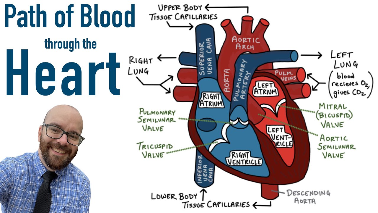
-Semilunar valves are located at the exit of the ventricles into the arterial system. There are two main semilunar valves: the pulmonary semilunar valve between the right ventricle and the pulmonary trunk, and the aortic semilunar valve between the left ventricle and the aorta.
How does the EKG represent the different stages of the cardiac cycle?
-The EKG represents the electrical activity of the heart. The P wave corresponds to atrial depolarization, the QRS complex indicates ventricular depolarization, and the T wave represents ventricular repolarization.
What is diastole and what happens during this phase?
-Diastole is the period of relaxation of the heart muscle. During this phase, the ventricles fill with blood coming from the atria as the AV valves are open, and the semilunar valves are closed.
What is the term used to describe the phase when both the atrioventricular and semilunar valves are closed?
-The phase when both the atrioventricular and semilunar valves are closed is known as the isovolumetric contraction phase.
What is the end diastolic volume (EDV) and when does it occur?
-The end diastolic volume (EDV) is the volume of blood in the ventricles just before the ventricles begin to contract. It occurs at the end of the ventricular filling phase when the atria have pushed the blood into the ventricles.
What is the significance of the dicrotic notch in the context of the cardiac cycle?
-The dicrotic notch is a brief rise in aortic pressure that occurs when the aortic semilunar valve closes, preventing backflow of blood into the aorta after ventricular ejection.
What is the first heart sound and what causes it?
-The first heart sound, also known as the 'Lub' or S1, is caused by the sudden closure of the atrioventricular valves at the beginning of ventricular systole.
What is the second heart sound and what causes it?
-The second heart sound, also known as the 'Dub' or S2, is caused by the closure of the aortic and pulmonary semilunar valves at the end of ventricular systole.
What is the isovolumetric relaxation phase and what happens during this phase?
-The isovolumetric relaxation phase is the period when the ventricles are relaxing and repolarizing after contraction. During this phase, the coronary arteries are filled, and the heart prepares for the next cycle by allowing the ventricles to refill with blood.
Outlines
😀 Introduction to the Cardiac Cycle
The video begins by introducing the cardiac cycle, which encompasses the mechanical events of blood flow through the heart's chambers, taking approximately 0.8 seconds. The discussion will cover the pressure differences in the atria and ventricles, the functioning of the atrioventricular (AV) valves and semilunar valves (SLV), and how these events correlate with the EKG. The speaker illustrates the pulmonary and aortic semilunar valves and describes the initial event of the cycle, which occurs during mid to late ventricular diastole, a period of relaxation when blood returns to the heart from various veins and the coronary sinus.
🏥 Ventricular Filling and Atrial Depolarization
The second paragraph explains the process of ventricular filling during the late phase of ventricular diastole. The atria fill with blood from the veins, increasing atrial pressure and causing the AV valves to open. Blood then passively flows into the ventricles due to gravity. The speaker emphasizes that 70-80% of blood enters the ventricles passively, with the remaining 20% pushed by atrial depolarization, which is triggered by the firing of the SA node. This atrial contraction is represented by the P wave on the EKG and signifies the end of ventricular diastole and the period of ventricular filling.
💓 Isovolumetric Contraction and Valve Dynamics
The third paragraph delves into the isovolumetric contraction phase, where the ventricles begin to contract but no blood is ejected yet. The ventricles' pressure rises as they squeeze, but it's still less than the pressure in the aorta and pulmonary trunk, keeping the semilunar valves closed. The speaker details the pressure differences, noting that the ventricles' pressure must exceed the arterial pressure for the semilunar valves to open. The atrioventricular valves close during this phase, producing the first heart sound, known as 'Lub' or S1.
🚀 Ventricular Ejection and Systolic Pressure Changes
In the fourth paragraph, the focus is on the ventricular ejection phase, where the ventricles continue to contract and their pressure exceeds that of the arteries, causing the semilunar valves to open. Blood is ejected into the pulmonary trunk and aorta. The AV valves remain closed due to the pressure differential. The speaker describes the end systolic volume (ESV), the remaining blood in the ventricles after contraction, and explains how the elastic arteries accommodate the increased pressure, leading to the closure of the semilunar valves and a brief rise in aortic pressure known as the dicrotic notch.
🔁 Isovolumetric Relaxation and Cardiac Cycle Completion
The final paragraph discusses the isovolumetric relaxation phase, where the ventricles start to relax and repolarize, leading to the second heart sound, 'Dub' or S2. The semilunar valves are closed, and the pressure in the ventricles decreases while the atria remain at low pressure. This phase marks the transition to diastole, where the ventricles will begin to fill with blood again, continuing the cardiac cycle. The speaker summarizes the entire cardiac cycle, emphasizing the continuous nature of the process and how it repeats every 0.8 seconds.
Mindmap
Keywords
💡Cardiac Cycle
💡Diastole
💡Atrial Pressure
💡Ventricular Pressure
💡Atrioventricular (AV) Valves
💡Semilunar Valves
💡EKG (Electrocardiogram)
💡End Diastolic Volume (EDV)
💡Isovolumetric Contraction
💡Ventricular Systole
💡End Systolic Volume (ESV)
Highlights
The cardiac cycle involves all mechanical events of blood flow through the heart chambers, averaging 0.8 seconds.
Differences in atrial and ventricular pressures, as well as arterial pressures, are key to understanding the cardiac cycle.
AV valves open when atrial pressure exceeds ventricular pressure, allowing blood to flow passively into the ventricles.
The tricuspid and bicuspid (mitral) valves are the AV valves that regulate blood flow between atria and ventricles.
Semilunar valves (pulmonary and aortic) close when arterial pressure is greater than ventricular pressure, preventing backflow.
The EKG's P wave represents atrial depolarization and contraction, pushing the remaining blood into the ventricles.
Isovolumetric contraction is the phase where the ventricles contract without ejecting blood, producing the first heart sound, 'Lub'.
Ventricular pressure must exceed arterial pressure for the semilunar valves to open and blood to be ejected into the arteries.
The closing of the semilunar valves produces the second heart sound, 'Dub', also known as S2.
The cardiac cycle includes phases of ventricular filling, contraction, ejection, and relaxation, correlating with specific EKG patterns.
The end systolic volume (ESV) is the remaining blood in the ventricles after contraction.
Elasticity of arteries allows them to accommodate increased pressure and recoil, distributing blood to the systemic and pulmonary circuits.
The dicrotic notch is a brief rise in aortic pressure when the semilunar valve closes, observable in a pressure graph.
The T wave on the EKG signifies ventricular repolarization and relaxation during the final phase of the cardiac cycle.
The cardiac cycle repeats every 0.8 seconds, ensuring continuous circulation of blood through the heart and body.
Transcripts
Browse More Related Video
The Ultimate Cardiac Cycle Video - Most Comprehensive on YouTube!

The Heart, Part 1 - Under Pressure: Crash Course Anatomy & Physiology #25
EKG/ECG Interpretation (Basic) : Easy and Simple!
Flow through the heart | Circulatory system physiology | NCLEX-RN | Khan Academy
Path of Blood Flow through the Heart | Step by step through every chamber, valve, and major vessel
Circulatory System and Pathway of Blood Through the Heart
5.0 / 5 (0 votes)
Thanks for rating: