Path of Blood Flow through the Heart | Step by step through every chamber, valve, and major vessel
TLDRThis educational video script delves into the intricacies of the heart's function, clarifying the myth that one should 'follow their heart' by explaining the heart's role as a muscular pump rather than a source of thoughts or feelings. It provides a detailed walkthrough of the blood flow through the heart's chambers and associated blood vessels, highlighting the importance of valves in unidirectional blood flow. The script also distinguishes between the oxygen-poor blood on the right side of the heart and the oxygen-rich blood on the left, concluding with a brief overview of the coronary arteries' role in supplying the heart muscle itself with blood.
Takeaways
- 🧡 The heart is primarily a muscular organ responsible for pumping blood throughout the body via contractions of cardiac muscle.
- 🔴 The heart is divided into four chambers: right atrium, right ventricle, left atrium, and left ventricle, each with specific roles in the blood circulation process.
- 💙 Blood flows from the atria to the ventricles and then outside the heart, with the right side handling low-oxygen blood and the left side handling high-oxygen blood.
- 🚦 Valves within the heart (tricuspid, pulmonary semilunar, mitral, and aortic semilunar) ensure one-way blood flow and prevent backflow.
- 💓 The right ventricle pumps blood through the pulmonary artery to the lungs, where it picks up oxygen and releases carbon dioxide.
- 💚 The oxygenated blood returns to the heart via the pulmonary veins, entering the left atrium and then the left ventricle.
- 💪 The left ventricle has thicker muscle than the right ventricle due to the greater distance it must pump blood throughout the body.
- 🔴🔵 The aorta, the body's largest artery, carries blood from the left ventricle to the rest of the body, including the coronary arteries that supply blood to the heart muscle itself.
- 🔄 Blood returns to the right atrium through the superior and inferior vena cavas after delivering oxygen to the body's tissues and picking up carbon dioxide.
- 📚 Understanding the path of blood flow is crucial for grasping the overall function of the circulatory system and diagnosing related medical conditions.
- 💓 The heart sounds people often associate with the heart's pumping action are actually the sounds of the heart valves closing.
Q & A
What is the primary function of the heart?
-The primary function of the heart is to pump blood throughout the body. It does this by contracting cardiac muscle, which moves blood through the heart's chambers and valves, and into the circulatory system.
How many chambers does the heart have, and what are their names?
-The heart has four chambers: the right atrium, right ventricle, left atrium, and left ventricle. The right atrium and ventricle handle deoxygenated blood, while the left atrium and ventricle handle oxygenated blood.
What is the role of valves in the heart?
-Valves in the heart ensure one-way blood flow. They open to allow blood to pass through and close to prevent backflow. Key valves include the tricuspid valve between the right atrium and right ventricle, the pulmonary semilunar valve between the right ventricle and pulmonary artery, the mitral valve between the left atrium and left ventricle, and the aortic semilunar valve between the left ventricle and aorta.
What is the path of low oxygen blood in the heart?
-Low oxygen blood enters the right atrium, passes through the tricuspid valve into the right ventricle, then through the pulmonary semilunar valve into the pulmonary artery, and travels to the lungs where it picks up oxygen and becomes high oxygen blood.
What is the path of oxygenated blood in the heart?
-Oxygenated blood from the lungs enters the left atrium through the pulmonary veins, passes through the mitral valve into the left ventricle, and is then pumped through the aortic semilunar valve into the aorta, from where it is distributed to the rest of the body.
Why does the left ventricle have thicker muscle than the right ventricle?
-The left ventricle has thicker muscle because it is responsible for pumping blood throughout the entire body, including to distant parts like the brain and extremities, which requires more force than the right ventricle's task of pumping blood to the nearby lungs.
What are the coronary arteries, and why are they important?
-The coronary arteries are small arteries that branch off from the aorta and supply blood to the heart muscle itself. They are crucial because any blockage in these arteries can lead to a heart attack, depriving the heart muscle of the oxygen it needs.
How does blood return to the heart after circulating through the body?
-Blood returns to the heart through the superior and inferior vena cavae. The superior vena cava collects blood from the upper body, while the inferior vena cava collects blood from the lower body. The blood then enters the right atrium to begin the circulation process again.
What is the significance of the heart's two-pump system with separate sides for low and high oxygen blood?
-The two-pump system allows for efficient oxygenation of blood. The right side collects deoxygenated blood from the body and sends it to the lungs for oxygenation. The left side receives oxygenated blood from the lungs and pumps it to the entire body, ensuring that all organs and tissues receive the oxygen they need for cellular respiration and energy production.
What is the role of the aorta in the circulatory system?
-The aorta is the largest artery in the body, originating from the left ventricle. It carries oxygen-rich blood away from the heart to the rest of the body through a series of branches, including the aortic arch branches that supply the upper body and the descending aorta that supplies the lower body.
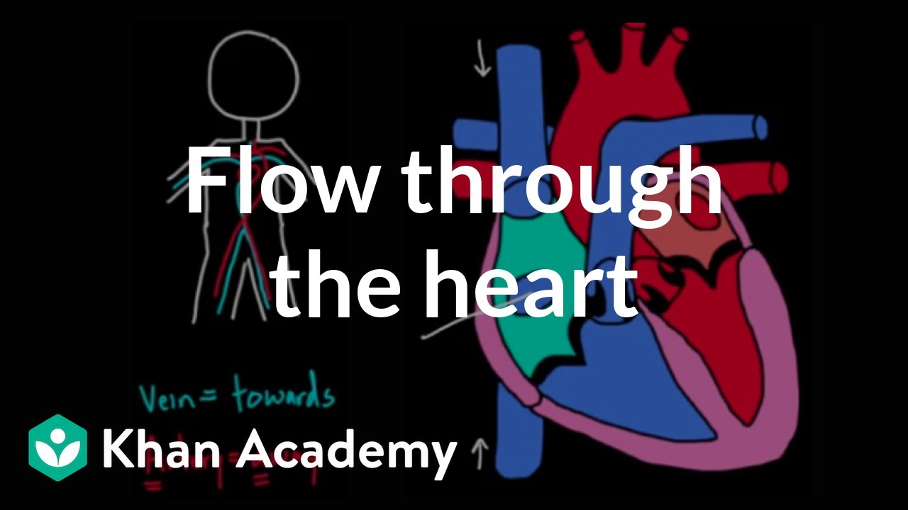
What does the color coding in the diagram represent?
-In the diagram, blue represents areas where blood is low in oxygen, and red represents areas where blood is high in oxygen. This color coding helps to visualize the path of deoxygenated and oxygenated blood through the heart and circulatory system.
Outlines
🧡 Understanding the Heart's Function and Blood Flow
This paragraph introduces the concept of following one's heart, clarifying that the heart is a muscular pump rather than a source of thoughts or feelings. It sets the stage for a detailed explanation of the heart's anatomy and the path of blood flow through its chambers and connected blood vessels. The speaker uses a whiteboard to diagram the heart, highlighting the four chambers, the valves that control blood flow direction, and the color-coding system to differentiate between oxygen-poor and oxygen-rich blood. The explanation emphasizes the heart's dual role in pumping low-oxygen and high-oxygen blood, and it begins to trace the path of blood from the right atrium to the lungs and back.
💙 The Journey of Oxygenated Blood
This paragraph continues the exploration of blood flow, focusing on the oxygenation process and the systemic circulation of blood. It describes the passage of blood from the right ventricle to the lungs via the pulmonary artery, where it picks up oxygen and releases carbon dioxide. The speaker explains the role of the pulmonary veins in returning oxygen-rich blood to the left atrium, and then its journey through the mitral valve into the left ventricle. The left ventricle's thicker muscle is highlighted as essential for pumping blood throughout the entire body. The paragraph also touches on the coronary arteries, which supply blood to the heart muscle itself, and the importance of these arteries in preventing heart attacks.
🔄 Completing the Circuit: Return of Deoxygenated Blood
The final paragraph wraps up the discussion on blood circulation by detailing the return of deoxygenated blood to the heart. It explains how blood flows from the body's tissues back to the right atrium through the superior and inferior vena cavas. The speaker reiterates that while the heart pumps blood out through arteries, it does not pump blood back into itself; instead, veins and gravity facilitate this process. The paragraph concludes with a recap of the entire blood flow pathway, emphasizing the importance of practice in understanding and remembering the sequence. Additionally, it provides a fun fact about the heart sounds being the result of valve closures, not the heart's pumping action.
Mindmap
Keywords
💡Heart
💡Blood Flow
💡Valves
💡Pulmonary Artery
💡Pulmonary Veins
💡Mitral Valve
💡Left Ventricle
💡Aorta
💡Superior and Inferior Vena Cava
💡Oxygenation
💡Cellular Respiration
Highlights
The heart is often metaphorically told to 'follow your heart', but it doesn't have thoughts or feelings; it's a pump.
The heart is primarily composed of muscle, which contracts to pump blood.
The heart has four chambers: right atrium, right ventricle, left atrium, and left ventricle.
Blood vessels are depicted in different colors to indicate oxygen levels, with blue for low oxygen and red for high oxygen.
The right side of the heart handles low oxygen blood, while the left side handles high oxygen blood.
Blood flows from the right atrium to the right ventricle through the tricuspid valve.
The pulmonary semilunar valve controls blood flow from the right ventricle to the pulmonary artery, heading towards the lungs.
In the lungs, blood picks up oxygen and releases carbon dioxide through the alveoli.
Oxygenated blood returns to the heart through the pulmonary veins, entering the left atrium.
The mitral or bicuspid valve allows blood to pass from the left atrium to the left ventricle.
The left ventricle has thicker muscle to pump blood throughout the entire body.
Blood exits the left ventricle through the aortic semilunar valve and into the aorta, the body's largest artery.
The aorta branches to supply blood to the upper and lower body through capillaries, where oxygen is delivered and carbon dioxide is collected.
Deoxygenated blood returns to the right atrium through the superior and inferior vena cava.
The heart's pumping action is not the source of its sound; it's the valves snapping shut.
Transcripts
Browse More Related Video
Flow through the heart | Circulatory system physiology | NCLEX-RN | Khan Academy

Heart Dissection GCSE A Level Biology NEET Practical Skills

The Heart, Part 1 - Under Pressure: Crash Course Anatomy & Physiology #25

Coronary circulation of the heart

The Human Heart | #aumsum #kids #science #education #children
Circulatory System and Pathway of Blood Through the Heart
5.0 / 5 (0 votes)
Thanks for rating: