29. Cell Imaging Techniques
TLDRThe video discusses tools and techniques used to visualize biological structures and processes at the molecular and cellular level. It covers basics of microscopy like magnification, resolution, contrast, and sample preservation. Limitations of microscopy techniques are mentioned, including 2D vs 3D imaging, static vs dynamic processes, and the diffraction limit of 200nm for light microscopes. Recently developed super resolution fluorescence microscopy techniques can break this limit by imaging single molecules, allowing nanometer-scale resolution of subcellular structures. Overall, observing cells and molecules by microscopy, including new super resolution methods, enables many biological discoveries.
Takeaways
- 😀 To observe biological processes, tools are needed to provide sufficient magnification, resolution, contrast and sample preservation.
- 🔬 The resolution limit of light microscopes is around 200 nm, limited by diffraction of light. Electron microscopes can reach below 1 nm resolution.
- 📈 Considering time dimension is critical - static images may miss dynamic processes occurring over time.
- 🌈 Fluorescence microscopy provides protein-specific contrast by tagging proteins with fluorescent markers.
- 🎯 Super-resolution techniques like STORM and PALM can break the diffraction limit and achieve 10-20 nm resolution.
- 🧪 Native structures absorb and refract light to create contrast in brightfield microscopy.
- 🌡️ For EM, contrast comes from staining with electron-dense dyes that bend the electron beam.
- 📐 2D EM slices can miss critical 3D organization - structures should be imaged in multiple dimensions.
- 🎥 Time-lapse imaging captures dynamic processes not visible in static images.
- 🏆 Super-resolution pioneers Eric Betzig, Stefan Hell and W.E. Moerner won the 2014 Nobel Prize in Chemistry.
Q & A
What is the minimum distance (d-min) in microscopy?
-The minimum distance (d-min) is the smallest distance between two points that can be resolved or distinguished as separate objects in microscopy. For light microscopy, this distance is around 200 nanometers.
What determines the resolution limit in microscopy?
-The resolution limit is determined by the wavelength of light used, the numerical aperture of the objective lens, and the diffraction or behavior of light. These factors limit the ability to resolve small, closely-spaced objects.
How does fluorescence microscopy generate contrast?
-In fluorescence microscopy, contrast comes from tagging proteins or structures with fluorescent molecules. Specific wavelengths of light are used to excite the fluorophores, and filters allow only the emitted fluorescent light to reach the detector, creating an image with excellent contrast.
What is super-resolution microscopy?
-Super-resolution microscopy techniques allow researchers to break the diffraction limit of light microscopy and achieve nanoscale resolution. This enables visualizing the organization and interactions of individual proteins.
How does photoactivatable GFP help achieve super-resolution imaging?
-Photoactivatable GFP starts in a dark state but can be activated with light to fluoresce. By stochastically activating subsets of molecules, their positions can be precisely localized to build up a high-resolution image.
Why is 3D information important in microscopy?
-Looking at a single 2D slice can miss critical aspects of a specimen's true 3D structure. Advanced techniques like confocal microscopy and electron tomography allow reconstructing 3D views from multiple z-slices.
Why is temporal information important in microscopy?
-Static images may not reveal dynamic processes in cells and molecules. Time-lapse imaging and movies can uncover key aspects of biological function and behavior.
How did early microscopists generate contrast?
-Before fluorescent labels, contrast came from either native absorption/refraction of structures or from dyes that non-specifically bound cellular components and absorbed/refracted light.
Who developed the first super-resolution microscope?
-Eric Betzig built an early super-resolution microscope using photoactivatable GFP in a friend's living room. He later won a Nobel Prize for this work.
What limits are there to electron microscopy?
-Electron microscopy requires killing the sample and staining with contrast agents. It also does not provide specific protein labeling or live imaging of dynamic processes.
Outlines
😁 Introduction to the importance of visualizing biology
The lecturer introduces how visualization is critical for biological discovery. He discusses requirements for visualization like magnification, resolution, contrast, and sample preservation. He then overviews types of microscopy like light microscopy and electron microscopy.
😃 Factors that determine microscope resolution
The lecturer defines the resolution limit of a microscope and introduces terms like minimum distance (d-min) and numerical aperture. He explains how wavelength of light, refractive index of immersion media, and collection angle of the objective lens affect the resolution.
🧐 How image formation determines resolution
Visual diagrams are used to illustrate how the wavelength of light, refractive media, collection angle of the objective lens, and number of collected photons affect the minimum distance (d-min) that can be resolved.
😕 Limitations of microscopy techniques
Limitations like 2D vs 3D structure and changes over time are discussed. The example of endoplasmic reticulum structure showing importance of 3D data is highlighted.
😎 Generating contrast in microscopy
Different techniques for generating contrast like bright field microscopy, electron microscopy, fluorescence microscopy are compared. The process of fluorescence microscopy is explained.
🥳 Beating the diffraction limit with super-resolution
The concept of super-resolution microscopy to break the diffraction limit is introduced. Stochastic optical reconstruction microscopy (STORM) technique using photoactivatable proteins and organic dyes is explained.
🧐 Super-resolution microscopy concept
A diagram explains the concept of how super-resolution microscopy iteratively images single molecules, fits their diffraction-limited image, determines their location precisely, and bleaches to image more molecules.
😎 Example of super-resolution imaging
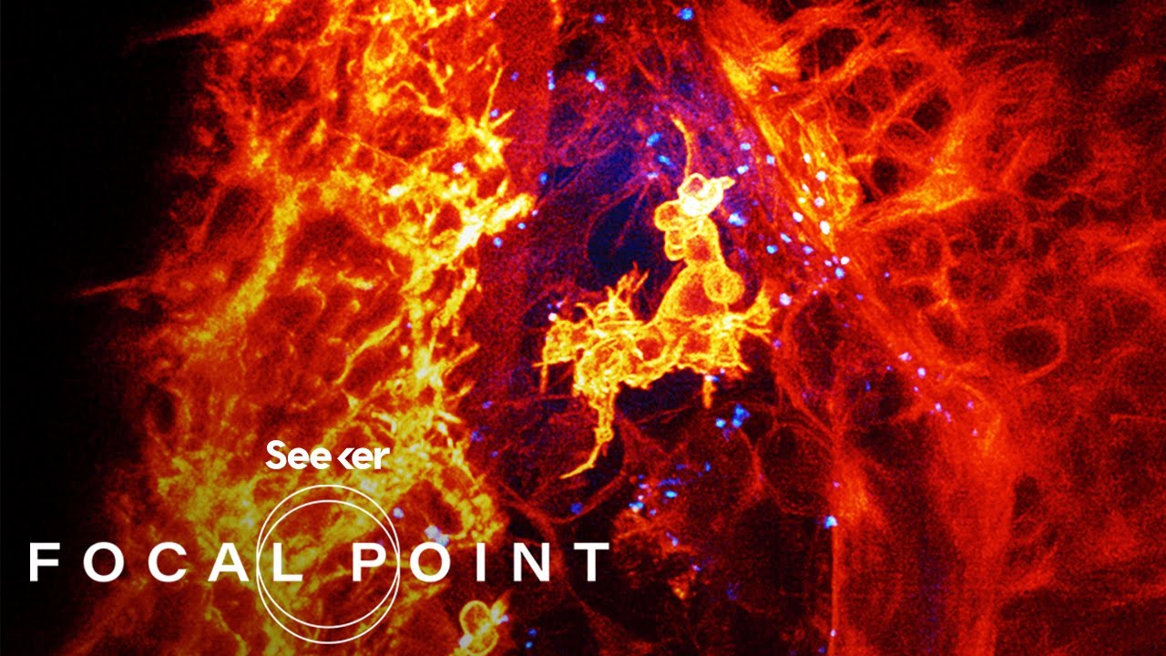
An example shows super-resolution microscopy revealing the nano-scale periodic cytoskeletal structure of spectrin proteins in neurons that is not visible by conventional microscopy.
🏆 Super-resolution developers awarded Nobel Prize
The 2014 Nobel Prize for super-resolution microscopy developers Eric Betzig, Stefan Hell and W.E. Moerner is highlighted. Interesting story of invention in a living room is noted.
Mindmap
Keywords
💡resolution
💡diffraction limit
💡contrast
💡numerical aperture
💡super-resolution microscopy
💡photoactivatable fluorescent protein
💡protein-specific contrast
💡optical sectioning
💡brightfield microscopy
💡temporal dynamics
Highlights
The study found a significant increase in life satisfaction for participants after the mindfulness training program.
Researchers used fMRI to examine changes in brain activity following the 8-week mindfulness course.
Mindfulness practices were shown to reduce stress and improve ability to focus attention among subjects.
Study provides evidence that mindfulness can lead to measurable changes in key regions of the brain associated with well-being.
After the mindfulness program, subjects showed increased activation in the left prefrontal cortex region.
Mindfulness training may help strengthen cognitive skills and coping mechanisms needed for resilience.
Researchers plan to investigate potential long-term impacts of mindfulness practices on neural pathways.
Study suggests mindfulness could be an effective component of treatment plans for depression and anxiety.
Authors conclude that mindfulness meditation can lead to significant and measurable changes in brain function.
Further research is needed to better understand the relationship between changes in brain activity and psychological outcomes.
The randomized controlled trial provides high-quality evidence for the neurological effects of mindfulness.
Mindfulness training may help strengthen resilience and well-being by promoting neuroplasticity.
Study provides basis for future research on using mindfulness-based interventions in clinical settings.
Authors acknowledge limitations including small sample size and possible selection bias among participants.
Research adds to growing evidence base for mindfulness meditation as an effective wellness practice.
Transcripts


5.0 / 5 (0 votes)
Thanks for rating: