Dental Anatomy | Occlusal Contacts | INBDE
TLDRIn this engaging Dental Anatomy video, Dr. Ryan delves into the concept of occlusal contacts, emphasizing the fundamental biological principle that structure dictates function. After reviewing the structure of permanent teeth, the focus shifts to their three-dimensional function. Dr. Ryan introduces three key planes: the coronal, axial, and sagittal, using memorable metaphors to aid understanding. The video explains the roles of functioning and non-functioning cusps and introduces the 'BULL rule' to identify cusps that should not be in contact during occlusion. The presenter then connects the coronal and axial planes to demonstrate how teeth interact, highlighting the facial and lingual ranges of occlusion. A simplified 'picket fence' diagram is used to illustrate tooth relationships in the sagittal plane, providing a valuable tool for board exam preparation. The video concludes with high-yield facts about ideal class 1 occlusion and a challenge for viewers to apply their knowledge, reinforcing the importance of foundational principles before advancing to more complex topics.
Takeaways
- 🦷 The core tenet of biology is the relationship between structure and function, which is crucial in understanding dental anatomy.
- 📏 There are three planes important for dental function: the coronal plane (left to right), the axial plane (horizontal split), and the sagittal plane (front to back).
- 👑 The term 'coronal plane' comes from the Latin word for crown, representing a plane similar to a crown or flat paper crown worn on the head.
- 🪓 The 'axial plane' is imagined as a woodcutter's ax chopping a tree horizontally, representing a horizontal plane.
- 🏹 The 'sagittal plane' is named after the Latin word for arrow, visualized as an arrow splitting the head into right and left halves.
- 📌 Functioning cusps of molars are centered over the opposing tooth for contact, while non-functioning cusps are taller and sharper to keep soft tissues away from the biting surfaces.
- ⚠️ The 'BULL rule' helps remember which cusps should not be in contact: Buckle (upper lingual) and Lower (facial) cusps.
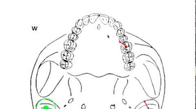
- 🔵🔴 The 'blue curve' represents the facial range of occlusion, and the 'magenta curve' represents the lingual range of occlusion when teeth are connected.
- 🧩 The 'picket fence diagram' is a simplified memory tool for visualizing how teeth fit together, useful for board exams.
- 📊 In a class 1 occlusion, the maxillary canine is the only tooth that contacts both an anterior and a posterior tooth.
- 🤝 All teeth occlude with two other teeth, except for the mandibular central incisor and the maxillary third molar, which only contact one other tooth.
- 📝 The picket fence diagram can be used to identify which teeth or cusps are in contact with specific areas of opposing teeth for diagnostic purposes.
Q & A
What is the core tenet of biology that Dr. Ryan discusses at the beginning of the video?
-The core tenet of biology discussed is the relationship between structure and function.
What are the three planes that are essential for understanding how teeth function together?
-The three planes are the coronal plane, the axial plane, and the sagittal plane.
How does Dr. Ryan suggest remembering the coronal plane?
-By associating it with a crown or Mickey Mouse ears, which are in the same plane as the coronal plane.
What is the 'bull rule' in the context of dental anatomy?
-The 'bull rule' stands for 'Buckle upper lingual, lower' and refers to the cusps that should not be in contact during occlusion.
What is the purpose of non-functioning cusps on teeth?
-Non-functioning cusps are taller and sharper, and their job is to hold the soft tissue, such as cheeks and tongue, away from the biting surfaces to prevent constant biting.
What does Dr. Ryan refer to as the 'blue curve' and 'magenta curve'?
-The 'blue curve' is the facial range of occlusion, and the 'magenta curve' is the lingual range of occlusion, representing the areas where the teeth come together when the mouth is closed.
What is the significance of the picket fence diagram in understanding dental occlusion?
-The picket fence diagram is a simplified memory tool that helps to visualize how the teeth fit together like puzzle pieces or the spokes on a gear, which is useful for board exam preparation.
How many vertical lines are drawn in the picket fence diagram?
-Nine vertical lines are drawn in the picket fence diagram.
What is a high-yield fact about the maxillary canine in an ideal class 1 occlusion?
-In an ideal class 1 occlusion, the maxillary canine is the only tooth that contacts both an anterior and a posterior antagonist.
How many other teeth does each tooth typically occlude with in an ideal occlusion?
-In an ideal occlusion, all teeth typically occlude with two other teeth, except for the mandibular central incisor and the maxillary third molar, which only contact one other tooth.
What is the purpose of the highlighted functioning cusp contacts in blue and magenta in the axial plane image?
-The highlighted functioning cusp contacts in blue and magenta in the axial plane image demonstrate the connection between the functioning facial and lingual cusps of the upper and lower teeth, respectively.
What is the importance of understanding the sagittal plane for the board exam?
-The sagittal plane is arguably the most important to memorize for the board exam as it provides a clear view of how the teeth fit together in a front-to-back orientation.
Outlines
🦷 Dental Anatomy: Occlusal Contacts and Planes
Dr. Ryan introduces the topic of occlusal contacts in the context of dental anatomy. He emphasizes the importance of understanding the relationship between tooth structure and function. The video covers three key planes of dental function: the coronal, axial, and sagittal planes. Dr. Ryan uses creative mnemonics to help viewers remember these planes, associating them with a crown, an ax, and a bow and arrow, respectively. He also discusses the roles of functioning and non-functioning cusps of teeth and introduces the 'BULL rule' to identify cusps that should not be in contact during occlusion to prevent dental issues.
📏 Understanding Cusp Contacts and Occlusion Curves
The video continues with a detailed look at the coronal and axial planes, highlighting the functioning cusp contacts and how they relate to the facial and lingual ranges of occlusion. Dr. Ryan explains the connection between the curvatures of the teeth and the occlusion curves, represented by blue and magenta lines. He then presents the 'picket fence diagram,' a simplified and memorable tool for visualizing tooth contacts and occlusion patterns, which is particularly useful for board exam preparation.
🧱 Picket Fence Diagram: A Study Aid for Occlusion
Dr. Ryan demonstrates how to construct the picket fence diagram, which represents the maxillary and mandibular arches and their corresponding teeth. The diagram is used to answer specific questions about tooth contacts, such as which cusp contacts the central fossa of the mandibular first molar or the distal marginal ridge of the mandibular first premolar. The video also reveals two high-yield facts for the board exam: the maxillary canine is the only tooth that contacts both an anterior and a posterior tooth in an ideal class 1 occlusion, and all teeth except the mandibular central incisor and the maxillary third molar occlude with two other teeth.
📝 Applying the Picket Fence Diagram to Board Exam Questions
The video concludes with practice questions that utilize the picket fence diagram to determine tooth contacts, such as identifying which teeth contact the distal marginal ridge of specific molars and premolars. Dr. Ryan provides answers and a trick for double-checking the answers by adding the tooth numbers, which typically sum up to 33, unless it's not a class 1 occlusion. He encourages viewers to practice these examples and to understand the foundational principles before moving on to more complex topics in subsequent videos.
Mindmap
Keywords
💡Occlusal Contacts
💡Coronal Plane
💡Axial Plane
💡Sagittal Plane
💡Functioning Cusps
💡Non-Functioning Cusps
💡Buckle Upper Lingual (BULL) Rule
💡Facial Range of Occlusion
💡Lingual Range of Occlusion
💡Picket Fence Diagram
💡Class 1 Occlusion
Highlights
The video discusses occlusal contacts in the context of dental anatomy, emphasizing the relationship between tooth structure and function.
Introduces three planes essential for understanding how teeth function together: coronal, axial, and sagittal planes.
Explains the coronal plane as the plane going from left to right, associated with the crown of a tooth.
Describes the axial plane as a horizontal slice dividing the tooth into superior and inferior halves, visualized with a woodcutter's ax.
Details the sagittal plane as a front-to-back plane, associated with the Latin word for arrow and the zodiac sign Sagittarius.
Discusses the functioning and non-functioning cusps of molars, their roles in occlusion, and the prevention of soft tissue damage.
Introduces the 'Buckle upper lingual, lower' rule to identify cusps that should not be in contact during occlusion.
Demonstrates how to visualize the connection between the coronal and axial planes through a diagrammatic representation.
Presents the facial range of occlusion (blue curve) and the lingual range of occlusion (magenta curve) for the upper and lower teeth.
Provides a simplified picket fence diagram to aid in memorizing tooth contacts and occlusion patterns.
Explains the use of the picket fence diagram for identifying specific tooth contacts during dental exams.
Highlights the maxillary canine as the only tooth in a class 1 occlusion that contacts both an anterior and a posterior tooth.
Mentions that all teeth except the mandibular central incisor and the maxillary third molar occlude with two other teeth.
Provides a method to check answers using the picket fence diagram, with a numerical pattern that often sums up to 33.
Encourages viewers to practice using the picket fence diagram to identify specific tooth contacts for exam preparation.
Advises understanding foundational principles before moving on to more complex topics such as working movement.
Invites viewers to like, subscribe, and support the channel for more content on Dentistry.
Transcripts


5.0 / 5 (0 votes)
Thanks for rating: